TomoSAXS X-ray tomography maps fibril-to-fibre mechanics in intact rat intervertebral discs
Authors combined phase-contrast CT and small-angle X-ray scattering to map nanoscale and microscale strain in intact discs under load; human discs were also imaged statically.
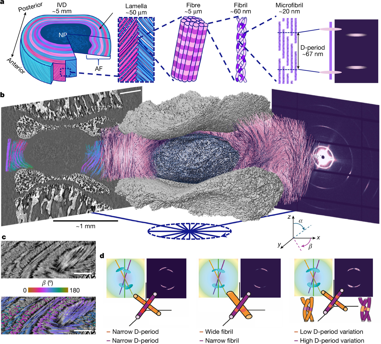
Researchers have described TomoSAXS, a full-field three-dimensional small-angle X-ray scattering tomography that maps mechanics from the fibril scale to the fibre scale across an intact tissue, according to a paper in Nature doi.org
Naturedoi.orgOpen source ↗. The method was applied to intact rat intervertebral discs (IVDs) under physiologically relevant loading and then extended to intact human IVDs under static conditions
doi.org
Naturedoi.orgOpen source ↗.
How the method works
TomoSAXS is a correlative, multiscale approach that integrates phase-contrast computed tomography (pCT) at the microscale with small-angle X-ray scattering (SAXS) at the nanoscale doi.org
Naturedoi.orgOpen source ↗. The pCT resolves the low-contrast fibre structure and, combined with digital volume correlation, gives volumetric deformation and strain at the microscale. Earlier fibre analysis of the IVD was generally limited to regions of interest smaller than 0.5 mm
doi.org
Naturedoi.orgOpen source ↗.
SAXS uses the collagen structure itself as a gauge. Fibrils of about 20–100 nm are formed of staggered tropocollagen molecules with a mean D-period of approximately 65–67 nm, which produces Bragg peaks at integer multiples of 1/D. Peak position along the radial axis reflects the D-period, peak width along the radial axis reflects its variation, and peak width along the circumferential axis reflects fibril diameter. Earlier 2D SAXS studies of the IVD were limited by the lack of 3D information doi.org
Naturedoi.orgOpen source ↗.
What the maps showed
The IVD consists of a hydrated nucleus pulposus surrounded by the annulus fibrosus. Its lamellae, tens of microns thick, contain collagen fibres 1–20 μm in diameter with alternating orientation between adjacent lamellae doi.org
Naturedoi.orgOpen source ↗. The authors report that intrafibrillar molecular pre-strain (D-period stagger) is lamellar textured and tightly correlated with microscale fibre strain
doi.org
Naturedoi.orgOpen source ↗.
Pre-strain was inversely related to fibril strain and its variability, which the authors describe as consistent with load-sharing through molecular unwinding doi.org
Naturedoi.orgOpen source ↗. Radial strain bridges and high-curvature zones at the annulus fibrosus–nucleus pulposus interfaces emerged as critical regulators of local mechanics
doi.org
Naturedoi.orgOpen source ↗.
The authors state that these findings reveal concerted fibril–fibre interactions that sustain mechanical equilibrium in the IVD, preserving elasticity and shape. They propose TomoSAXS as a platform for other biological and synthetic fibrous materials, naming ageing and disease, therapeutic evaluation, and the design of bio-based and bio-inspired materials as areas of application doi.org
Naturedoi.orgOpen source ↗. IVD injury and age-related degeneration are described as a key driver of back pain, the leading global cause of years lived with disability
doi.org
Naturedoi.orgOpen source ↗.