PGTI method reconstructs proton velocity and range from prompt-gamma timing in head phantom
Authors combined eight TIARA detector modules to reconstruct tissue sequence and proton range in a head phantom irradiated with 100 MeV protons; the range differed by about 1 mm.
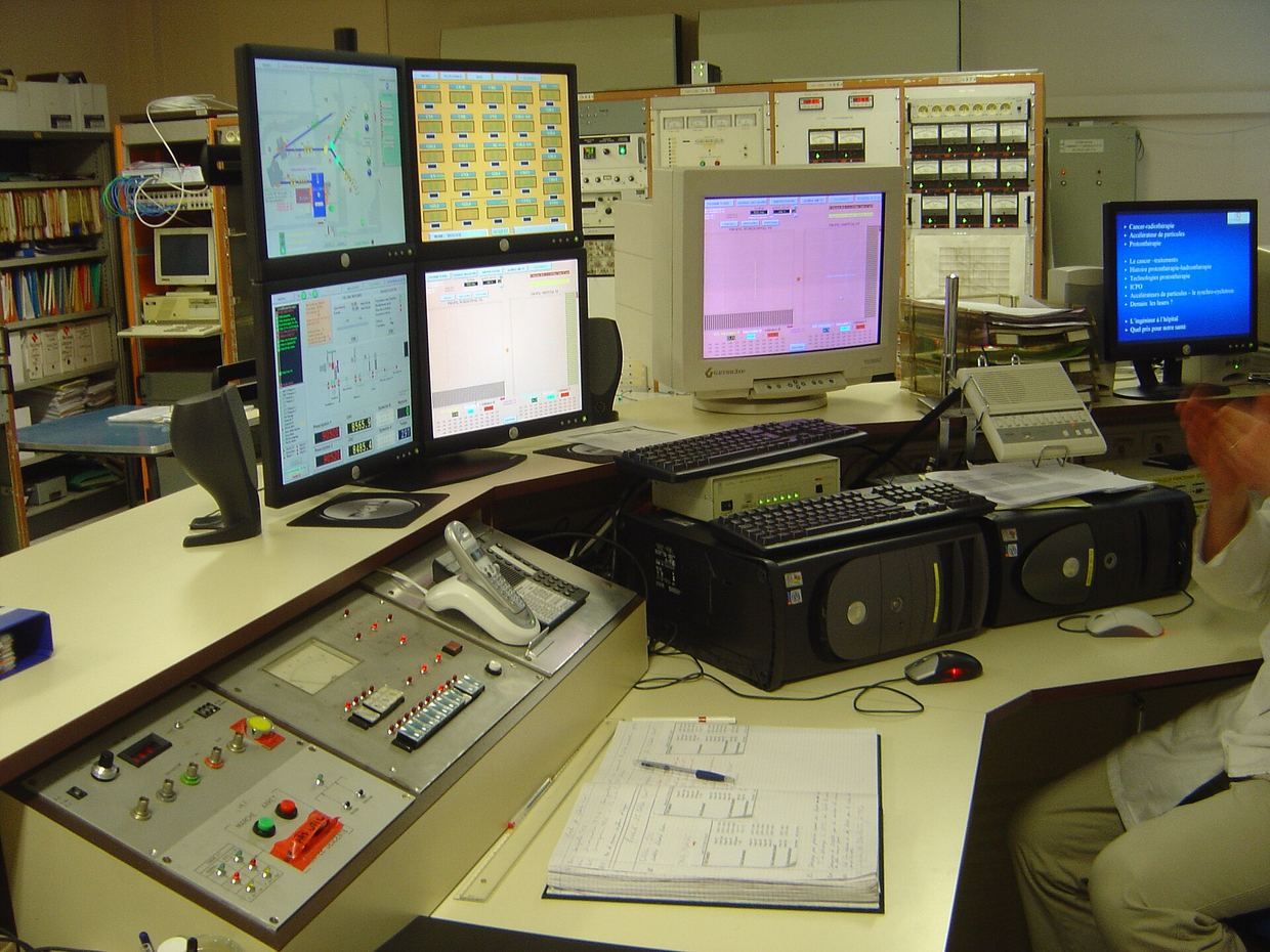
A collaboration working on the TIARA detector system has described Prompt-Gamma Time Imaging (PGTI), an iterative reconstruction method for monitoring proton beams. The work was published as an arXiv · preprint and has not been peer reviewed. The authors tested it on an anthropomorphic RANDO head phantom irradiated with 100 MeV protons at the ProteusOne accelerator of Centre Antoine Lacassagne arxiv.org
arXiv · препринтarxiv.orgOpen source ↗.
What the method does
Proton therapy relies on knowing the proton range inside the patient. It can be affected by anatomical changes, setup uncertainties and errors in converting CT numbers into stopping power. Prompt gamma rays are emitted almost instantly after nuclear interactions and leave the body with little interaction, so their detection can be used for in vivo verification.
Conventional prompt-gamma timing is hard to apply to several detectors at once, because the time of flight depends on the detector position. PGTI accounts for the detector geometry and jointly estimates two quantities from time-of-flight data: the prompt-gamma emission distribution and the proton velocity profile arxiv.org
arXiv · препринтarxiv.orgOpen source ↗. According to the authors, velocity is directly related to stopping power, so it gives access to tissue composition along the beam and a first-order estimate of the delivered dose. The approach also removes the dependence on prior anatomical assumptions used in an earlier reconstruction formalism.
Experiment with the phantom
TIARA combines a fast scintillator beam monitor with compact gamma detector modules (15 × 15 × 20 mm³) based on pure Cherenkov radiators. The current prototype has eight modules; a final clinical version is expected to have up to thirty. Here the modules were placed symmetrically on both sides of the phantom, about 15 cm from its center, with the beam directed at a sinus. The accelerator ran in single-proton regime, about one proton per bunch.
Two geometries imitated a change between planning and delivery: a sinus filled with ultrasound gel and an empty sinus. The authors recorded 6.9 × 10³ events for the empty configuration and 6.2 × 10³ for the filled one. They state this corresponds to the statistics a 30-module system would collect from slightly more than two spots of 10⁷ protons at 100 MeV. A separate run without the phantom was used to subtract background.
The reconstructed material sequence agreed with the reference CT. Ten bins had a misclassified material, yet the reconstructed proton range differed from the expected value by about 1 mm arxiv.org
arXiv · препринтarxiv.orgOpen source ↗. The authors report that the method also identified the anatomical change. They describe this as the first demonstration of PGTI under realistic conditions and as a validation of TIARA in a pseudo-clinical environment.