Review in Med Research compares four organoid coculture methods for modelling the tumor microenvironment
A review in Med Research describes coculture systems that add fibroblasts, immune cells, flow and vasculature to cancer organoids, aiming at better preclinical drug testing.
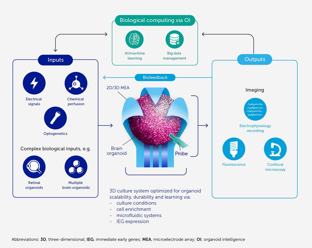
A review by Yang Z. and co-authors, published in 2026 in Med Research, summarizes organoid coculture models for cancer research and immunotherapy doi.org
Med Researchdoi.orgOpen source ↗. According to the authors, three-dimensional cancer organoids grown in vitro closely reproduce the phenotypic and genetic features of the parental tumor tissue. This makes them used for cancer research, drug screening and personalized medicine
doi.org
Med Researchdoi.orgOpen source ↗.
The authors note that traditional organoid cultures lack key components of the tumor microenvironment (TME). These include cancer-associated fibroblasts, immune cells, vascular networks, and dynamic physicochemical cues such as fluid flow and oxygen gradients doi.org
Med Researchdoi.orgOpen source ↗. As a result, it is harder to study tumor–stroma crosstalk, model responses to immunotherapy and predict clinical efficacy, particularly for agents that act through intercellular interactions
doi.org
Med Researchdoi.orgOpen source ↗.
Matrigel and air–liquid interface cocultures
Submerged Matrigel coculture allows specific TME components to be reconstituted systematically, so that defined cell–cell interactions can be dissected doi.org
Med Researchdoi.orgOpen source ↗. Air–liquid interface culture preserves the native, unmanipulated TME and retains resident immune cells and fibroblasts. According to the review, this suits studies of immune infiltration and checkpoint blockade
doi.org
Med Researchdoi.orgOpen source ↗.
Microfluidic chips and 3D bioprinting
Microfluidic culture and organoid-on-a-chip platforms provide dynamic control over fluid flow, oxygen gradients and shear stress. The authors state that this overcomes the static nature of traditional cultures and enables vascularization, perfusion modeling and high-throughput drug screening doi.org
Med Researchdoi.orgOpen source ↗. Three-dimensional bioprinting is described as offering high spatial precision in building multilayered heterogeneous tumor architectures, including vascular networks and tumor–stroma interfaces
doi.org
Med Researchdoi.orgOpen source ↗.
The review also covers applications of these systems in immunotherapy optimization, adoptive cell therapy and personalized medicine. It analyzes the advantages and limitations of each model and outlines perspectives for developing more physiologically relevant preclinical platforms doi.org
Med Researchdoi.orgOpen source ↗. It is a literature synthesis and does not report a new experiment.