Kanazawa University films Smc5/6 complex binding DNA at submolecular resolution in vitro
High-speed AFM resolved individual domains of yeast Smc5/6 on DNA, showing two binding modes and tethering of two segments. The experiments used purified proteins outside cells.
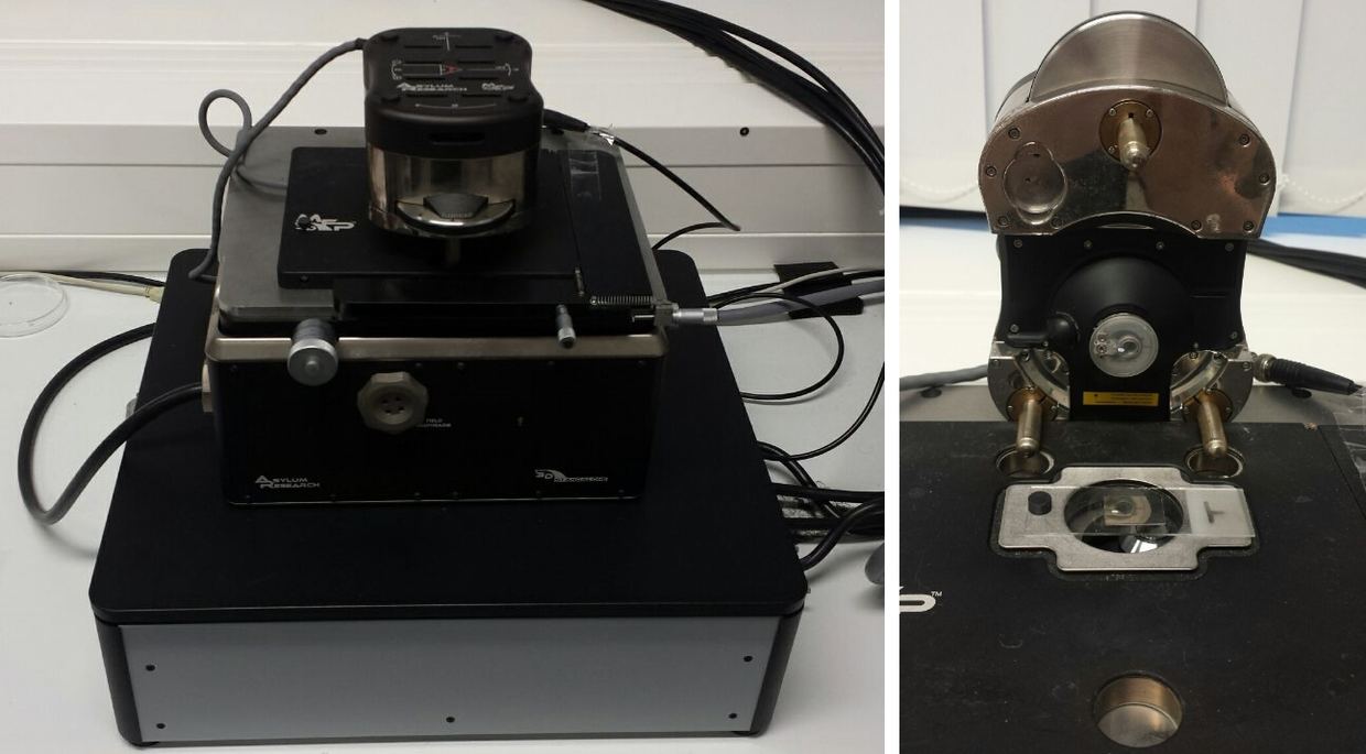
Researchers at Kanazawa University and the National Institute of Genetics in Japan used high-speed atomic force microscopy (HS-AFM) to follow the Smc5/6 complex on DNA. The study, by Kenichi Umeda, Yumiko Kurokawa, Noriyuki Kodera and Yasuto Murayama, appears in Nature Communications 17, 10398 (2026) doi.org
Nature Communicationsdoi.orgOpen source ↗. The images resolve individual domains of the complex at submolecular scale
doi.org
Nature Communicationsdoi.orgOpen source ↗.
How Smc5/6 holds DNA
SMC complexes are ring-shaped ATPases that hold distinct DNA segments together and regulate chromosome structures and functions. Their dynamic mechanisms of DNA binding and processing have remained poorly understood doi.org
Nature Communicationsdoi.orgOpen source ↗. The team studied purified Smc5/6 from budding yeast under controlled laboratory conditions.
According to the authors, ATP-bound Smc5/6 stably binds DNA via the ATPase head domain. After ATP hydrolysis, the DNA is entrapped within the SMC compartment and positioned around the opposite hinge domain doi.org
Nature Communicationsdoi.orgOpen source ↗. The two states correspond to the head-binding and hinge-binding modes shown in the images.
The complex also adopted two shapes. In one, its two long arms lay close together and resembled the letter I. In the other, the arms separated into an open structure resembling the letter O. ATP influenced the transition between them, and ATP binding promoted the open configuration.
Tethering and compaction
Smc5/6 topologically entraps two DNA segments together, a process known as DNA tethering doi.org
Nature Communicationsdoi.orgOpen source ↗. It also stabilizes DNA twist structures and promotes DNA compaction
doi.org
Nature Communicationsdoi.orgOpen source ↗. When the complex interacted with twisted DNA, the DNA occupied a smaller area than DNA without the complex.
"By observing individual Smc5/6 structures as they interacted with DNA, we could see how changes in their shape are associated with different ways of capturing and holding DNA," said Kenichi Umeda of Kanazawa University.
The authors describe the results as a visual demonstration of how an SMC complex uses its ring architecture to produce distinct DNA binding modes doi.org
Nature Communicationsdoi.orgOpen source ↗.
Limits of the experiment
The work used purified Smc5/6 and DNA outside living cells. The images therefore show molecular interactions under controlled conditions and do not directly establish every step of Smc5/6 activity inside a cell. The study belongs to fundamental molecular biology and does not demonstrate a medical treatment or clinical application.