Neanderthal heel bones were shorter, wider and taller than in Homo sapiens, 3D study finds
Researchers compared 7 Neanderthal and 64 Homo sapiens calcanei with 3D geometric morphometrics. The shapes suggest a habitually pronated foot and heavy load on the rearfoot.
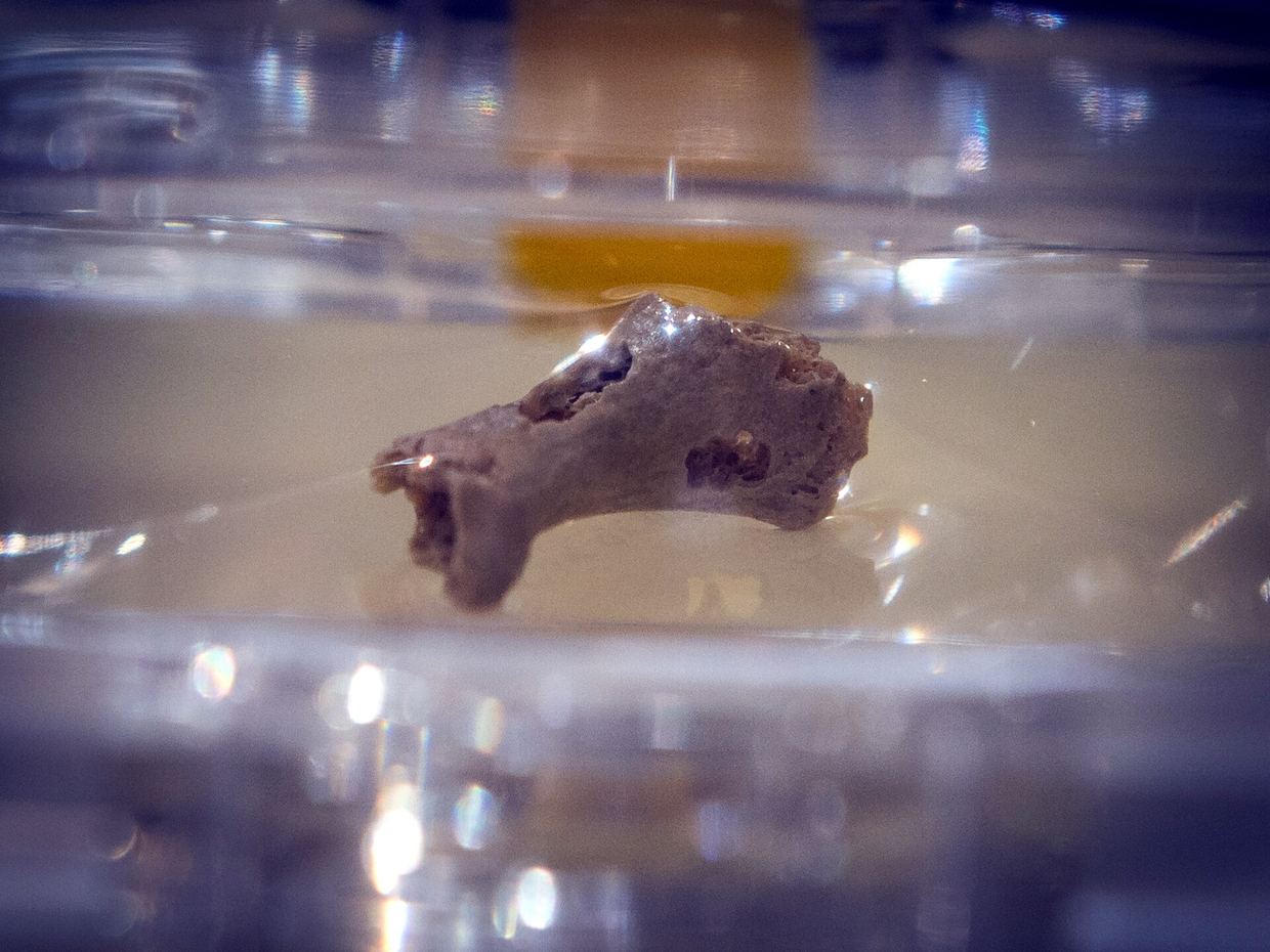
A team from the University of Bologna and partner institutions analysed the three-dimensional external shape of the calcaneus, the heel bone, in 7 Neanderthals and 64 Homo sapiens. The results were published in the Journal of Human Evolution doi.org
Journal of Human Evolutiondoi.orgOpen source ↗. The Homo sapiens sample ranged from Upper Pleistocene hunter-gatherers and Iron Age farmers to a post-industrial urban population from the Documented Human Osteological Collection of the Certosa Cemetery of Bologna. The work was led by Rita Sorrentino and coordinated by Maria Giovanna Belcastro.
Calcaneus shape and size
The authors applied geometric morphometrics to digital models obtained with laser scans and computed tomography. They examined the whole bone and its subregions: the talar facets, the cuboid facet and the tuberosity doi.org
Journal of Human Evolutiondoi.orgOpen source ↗. Compared with H. sapiens, Neanderthal calcanei were shorter, wider and taller. According to the authors, these traits likely favoured load-bearing over the rearfoot and high mobility
doi.org
Journal of Human Evolutiondoi.orgOpen source ↗.
Similar but less pronounced traits appeared in highly mobile, unshod modern humans. Sedentary post-industrial populations had more gracile calcanei, which the authors link to footwear and reduced activity doi.org
Journal of Human Evolutiondoi.orgOpen source ↗. Belcastro said the robust heel reflects the overall Neanderthal body structure, with high muscular and bone mass and intense physical activity.
Foot posture and arch
Neanderthals also kept ancestral features, such as a shorter sinus tarsi doi.org
Journal of Human Evolutiondoi.orgOpen source ↗. The shape of their subtalar joint and the orientation of the calcaneocuboid joint suggest a habitually pronated foot posture
doi.org
Journal of Human Evolutiondoi.orgOpen source ↗. The lateral plantar process was reduced compared with H. sapiens, which implies differences in plantar ligament attachment and foot arch support
doi.org
Journal of Human Evolutiondoi.orgOpen source ↗.
Differences in the cuboid facet and tuberosity point to alternative strategies of foot stabilization and force transmission through the proximal tarsal joints. The authors link these to a different expression of the longitudinal arch, adapted to Neanderthal body mass and a high-activity lifestyle doi.org
Journal of Human Evolutiondoi.orgOpen source ↗. In the university's summary, this is described as a flat foot that reduced impact stress under heavy loads and gave stability on rough terrain.
The university release also notes that rigid-soled shoes tend to limit foot inversion and eversion, and treats the gracile post-industrial heel as a sign of reduced biomechanical stimulation. Belcastro said the results may help dialogue between evolutionary anthropology and clinical orthopaedics.