Edinburgh team: Prototaxites taiti fossils from Rhynie chert differ from fungi in chemistry and structure
Fossils of Prototaxites, a Devonian organism up to eight meters tall, differ from fungi, so the authors place it in an entirely extinct lineage.
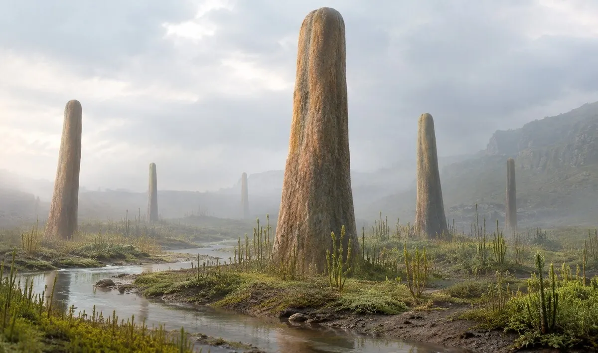
A team from the University of Edinburgh led by Corentin C. Loron, Laura M. Cooper and Alexander J. Hetherington compared the organization and molecular composition of Prototaxites taiti with those of fungi. The fossils come from the 407-million-year-old Rhynie chert in Scotland, and the results were published in Science Advances. science.org
Science Advancesscience.orgOpen source ↗ According to the authors, the fossils were chemically distinct from contemporaneous fungi and structurally distinct from all known fungi.
Protaxites is the first giant organism on the land surface, known from columnar fossils up to eight meters tall from the Early Devonian. Its systematic affinity has been debated for more than 165 years. Two hypotheses remained viable: a fungus, or a member of an entirely extinct lineage. The new data cast doubt on the fungal affinity and point to an extinct eukaryotic lineage.
Rhynie chert specimens
The work centered on three specimens of P. taiti, including a well-preserved one catalogued as NSC.36. In the rock block it is about 6.9 cm long and 5.6 cm thick at its widest; both ends are missing. This is the smaller species, not one of the meter-high specimens, so the sample did not measure the size of the giants. National Museums Scotland accepted one of the studied fossils into its collection.
The Rhynie chert preserves several groups of organisms under comparable conditions, which allows their chemical composition to be compared directly. An earlier interpretation had linked supposed reproductive structures of this species to ascomycete fungi. On re-examination, the team found that the fragments were not demonstrably connected to material that clearly belongs to Prototaxites.
Tube types and medullary spots
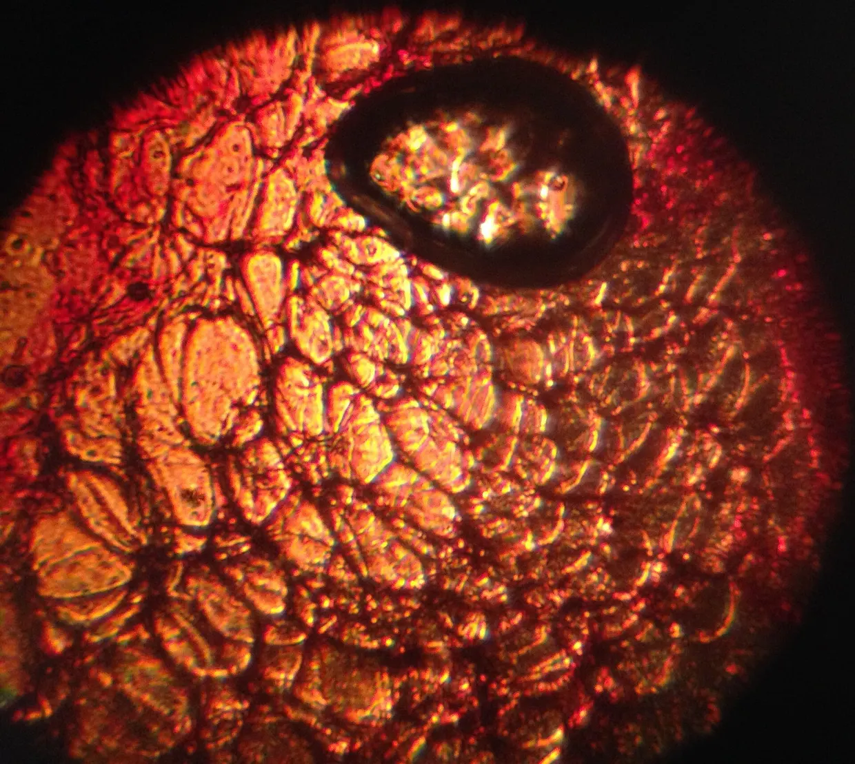
Under the microscope, P. taiti is a network of several types of tubes. Some are very thin and have cross-walls, others are wider and lack them, and others have ring-shaped wall thickenings that are unusual in this arrangement in known fungi. Inside, the researchers also found small dark areas called medullary spots.
Laser microscopy and a three-dimensional reconstruction showed how tubes branched within these spots. Different tube types merged into a tightly connected network without the order typical of fungal tissues. Hetherington noted that comparable structures are absent from anatomical descriptions of living fungi. The network may have exchanged water, gases or nutrients between body regions, but this function was inferred from its shape and not measured directly. The study does not resolve what Prototaxites was; it narrows the options.