UT Southwestern's miOPM light-sheet microscope swaps oil, water and air objectives
A team led by UT Southwestern described a light-sheet microscope that keeps diffraction-limited imaging with oil, water and air objectives, from subcellular dynamics to cleared tissue.
A team led by UT Southwestern Medical Center in Dallas has developed the multi-immersion oblique plane microscope (miOPM), a light-sheet fluorescence platform for 3D imaging of biological samples. The work was published in Nature Methods and is described in the nature.com paper; UT Southwestern announced it on 5 November 2026 nature.com
nature.comnature.comOpen source ↗. The corresponding author is Reto Fiolka, Associate Professor in the Lyda Hill Department of Bioinformatics and of Cell Biology.
According to the authors, light-sheet microscopy has split into variants, each optimized for a narrow range of samples or imaging regimes. High-resolution variants use a detection objective paired with a custom illumination objective, and these objectives cannot readily be swapped nature.com
nature.comnature.comOpen source ↗. As a result, systems suited to single-cell dynamics are poorly suited to whole embryos, and cleared tissues often need their own configurations
nature.com
nature.comnature.comOpen source ↗.
One objective, several immersion media
Oblique plane microscopy uses a single objective for illumination and detection. Until now, changing that objective meant changing the whole downstream optical train, in effect building a new microscope nature.com
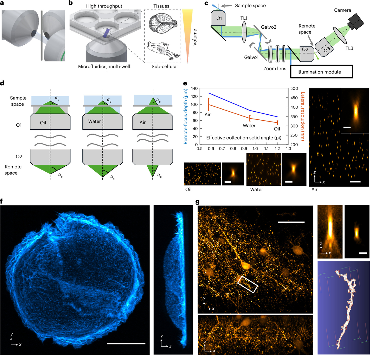
nature.comnature.comOpen source ↗. In miOPM, oil, water and air objectives are interchangeable, and the authors report diffraction-limited performance across a refractive index range of 1.33 to 1.51
nature.com
nature.comnature.comOpen source ↗.
The platform is compatible with standard sample mounting, current clearing protocols and high-throughput 3D imaging, the authors say nature.com
nature.comnature.comOpen source ↗. Lateral resolution was estimated from images of fluorescent nanospheres in agarose with air, water and oil objectives
nature.com
nature.comnature.comOpen source ↗.
Demonstrated samples
With an oil immersion objective, the team imaged a SU8686 cell labeled with Tractin-mRuby. With a silicone oil objective, they imaged a CLARITY-cleared mouse brain section labeled for Thy1-eGFP neurons and segmented an axon from it nature.com
nature.comnature.comOpen source ↗. Images released with the announcement also show carbohydrate-rich molecules in mouse lung tissue, mitochondria and mitochondrial DNA in a HeLa cell, and actin filaments in a human pancreatic cancer cell.
The authors say the design could let laboratories cover more applications with one instrument and reduce the need for several specialized microscopes. Fiolka said the work makes light-sheet fluorescence microscopy more versatile and user-friendly and extends what a single instrument can do in sample coverage and imaging applications.