University of Houston team builds handheld prototype for label-free mid-infrared chemical imaging of tissue
A handheld fiber-optic probe maps the molecular composition of tissue without stains; in lab tests on thin preserved slices it matched a benchtop instrument.
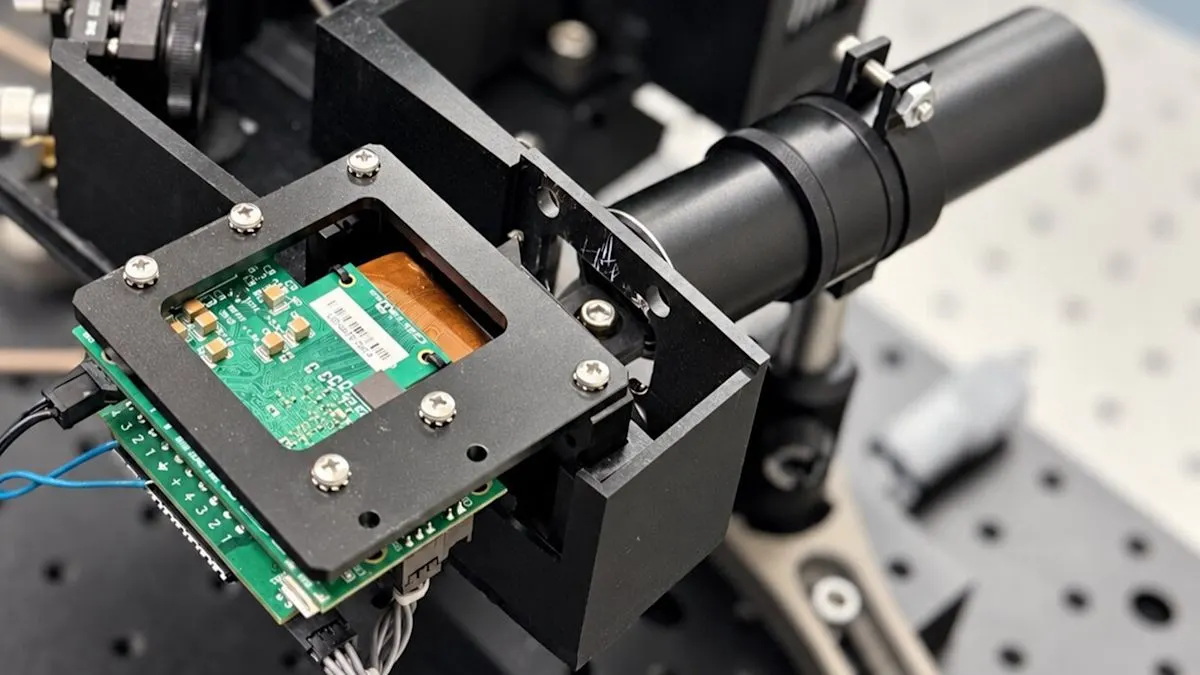
A team at the University of Houston led by Rohith Reddy has built a handheld version of photothermal mid-infrared spectroscopic imaging (MIRSI), according to a paper in Optica optica.org
Opticaoptica.orgOpen source ↗. Until now such platforms were bulky benchtop instruments confined to controlled laboratories. The authors say this is, to their knowledge, the first time the method has been used outside the lab. The scanner probe is 20 cm (8 inches) long, and the bone marrow samples came from a pathologist at MD Anderson Cancer Center.
How the probe works
Molecules absorb mid-infrared light at wavelengths set by their chemical bonds, so proteins, fats and DNA each have a characteristic pattern. The system aims two lasers at one spot. A mid-infrared laser fires pulses 1,000 times a second, and where molecules absorb the light the spot warms slightly. A red laser detects the resulting change, and sweeping the spot across a sample gives a map of a chosen chemical without any stain.
The hard part was making both beams hit exactly the same point. The team sent the infrared light through a glass fiber made for those wavelengths and replaced lenses with mirrors; a curved mirror at the tip focuses both beams. The lasers and electronics sit in a base unit of less than 1.5 square feet (0.14 square meters), while commercial instruments of this kind occupy more than 9 square feet (0.84 square meters) of optical table. The scanner offers configurable scanning modes: a moving mirror inside the probe or a stage that shifts the sample.
What the tests showed
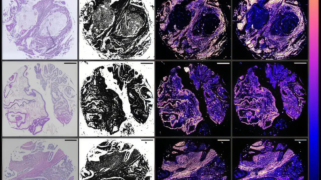
On a test chart the probe resolved details about 2 micrometers across, and its images closely matched a commercial benchtop instrument. On thin slices of cervical cancer from two patients and ovarian cancer from two others, handheld and benchtop images correlated at 0.90 at a coarse level of detail. In ovarian tissue, chemical patterns agreed with a standard laboratory infrared measurement at 0.935 optica.org
Opticaoptica.orgOpen source ↗.
In bone marrow, the unstained scan showed collagen around a blood vessel, matching the blue in an adjacent stained slice. In mouse kidney tissue it showed a glomerulus with chemical detail absent from the red-laser image alone.
Every sample was a thin, preserved slice on a slide; living or freshly excised tissue was not imaged. The team has not yet tested whether the scans help distinguish cancer from healthy tissue. Reddy called the platform a research prototype and said it could eventually allow tissue assessment during cancer surgery. The researchers plan to widen the infrared range and speed up scanning, and repeatability, safety and diagnostic performance still have to be tested in clinical conditions.