Choroid plexus microstructure altered in mild small vessel disease, MRI study of 139 patients
In a clinical MRI study, choroid plexus microstructural changes appeared in mild cerebral small vessel disease without enlargement of the structure, alongside altered perivascular fluid markers.
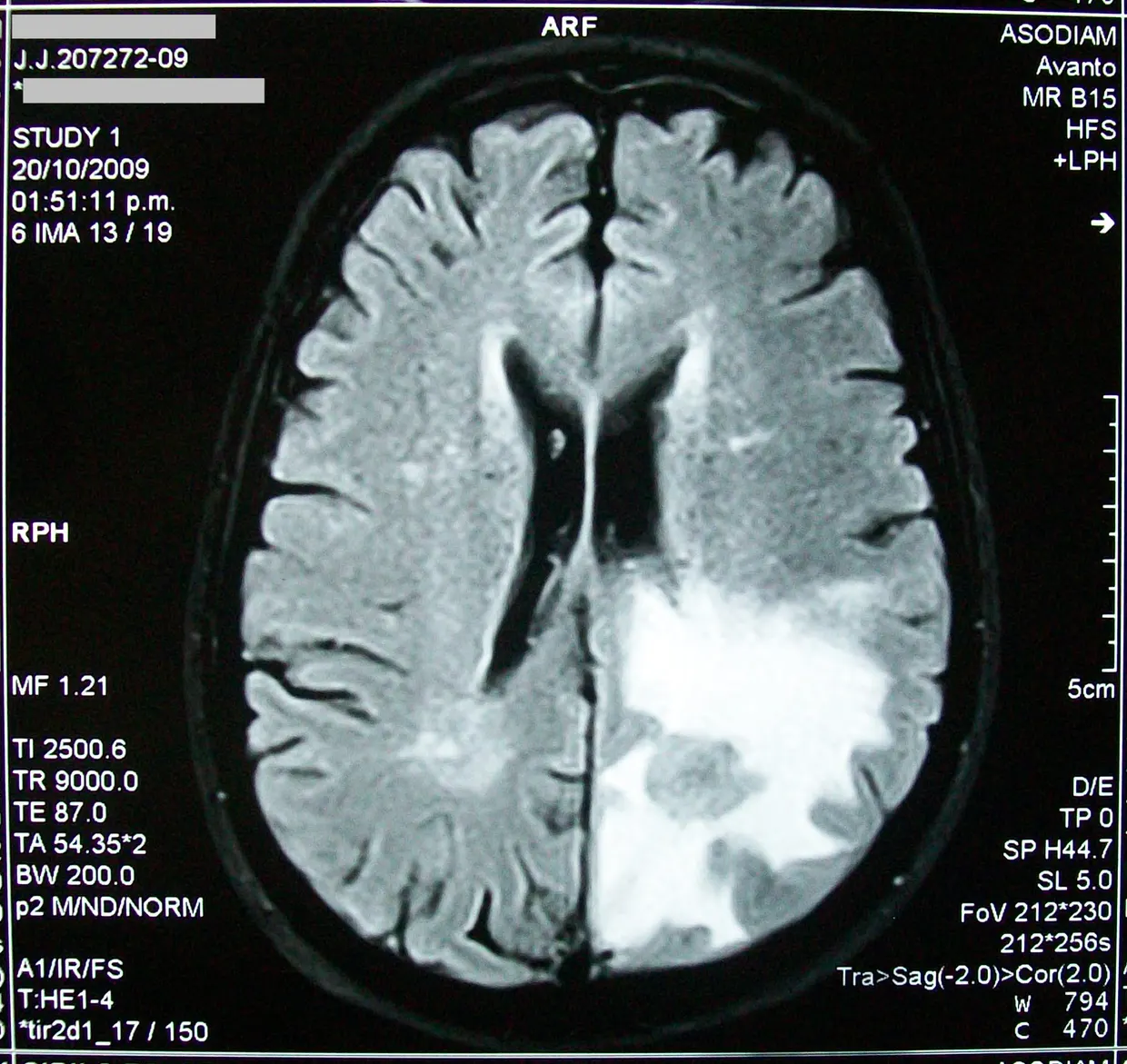
A research team reported in Brain Informatics an imaging study of the choroid plexus (CP) in cerebral small vessel disease (CSVD). The study included 139 CSVD patients and 52 healthy controls. According to the authors, microstructural injury of the CP is present in mild CSVD even when the CP is not enlarged biomedcentral.com
Brain Informaticsbiomedcentral.comOpen source ↗.
Choroid plexus and perivascular clearance
The CP is the main blood-cerebrospinal fluid barrier and synthesizes a substantial share of cerebrospinal fluid (CSF). Its dysfunction may impair CSF turnover and perivascular glymphatic clearance, but it was unclear whether CP microstructural injury relates to CSVD-related brain alterations and cognitive performance. Earlier imaging work mostly tracked CP enlargement, which cannot show early cellular changes.
CP microstructure was assessed by mean apparent propagator (MAP) diffusion imaging and by χ-separation susceptibility mapping. Perivascular clearance was characterized with three markers: the diffusion tensor image-along perivascular space (DTI-ALPS) index, the basal ganglia free-water fraction (FW-BG) and the basal ganglia perivascular space volume fraction (PVSVF-BG).
Results and mediation analysis
Compared with controls, CSVD patients showed CP microstructural abnormalities and altered perivascular clearance markers. A random-forest model identified these features as predictors of CSVD severity (OOB-AUC = 0.755, 95% CI: 0.641–0.836) biomedcentral.com
Brain Informaticsbiomedcentral.comOpen source ↗. Overt CP enlargement was found only in the severe subgroup, while microstructural changes were already present in mild cases.
Mediation analysis showed that the perivascular clearance markers significantly mediated the associations between CP alterations and structural brain injury (average causal mediation effect, ACME = −110.118 to 80.121, FDR-p < 0.05). PVSVF-BG was the only glymphatic imaging metric that mediated the links of both CP volume and CP susceptibility with cognitive performance (ACME = −142.474 to 67.351, FDR-p < 0.05).
The authors describe the CP-glymphatic axis as a candidate pathway for further longitudinal and mechanistic investigation. The work reports associations measured by imaging and does not establish causation.