Bielefeld and Vienna teams combine SIM and FLIM, reaching 156 nm resolution on bone cancer cells
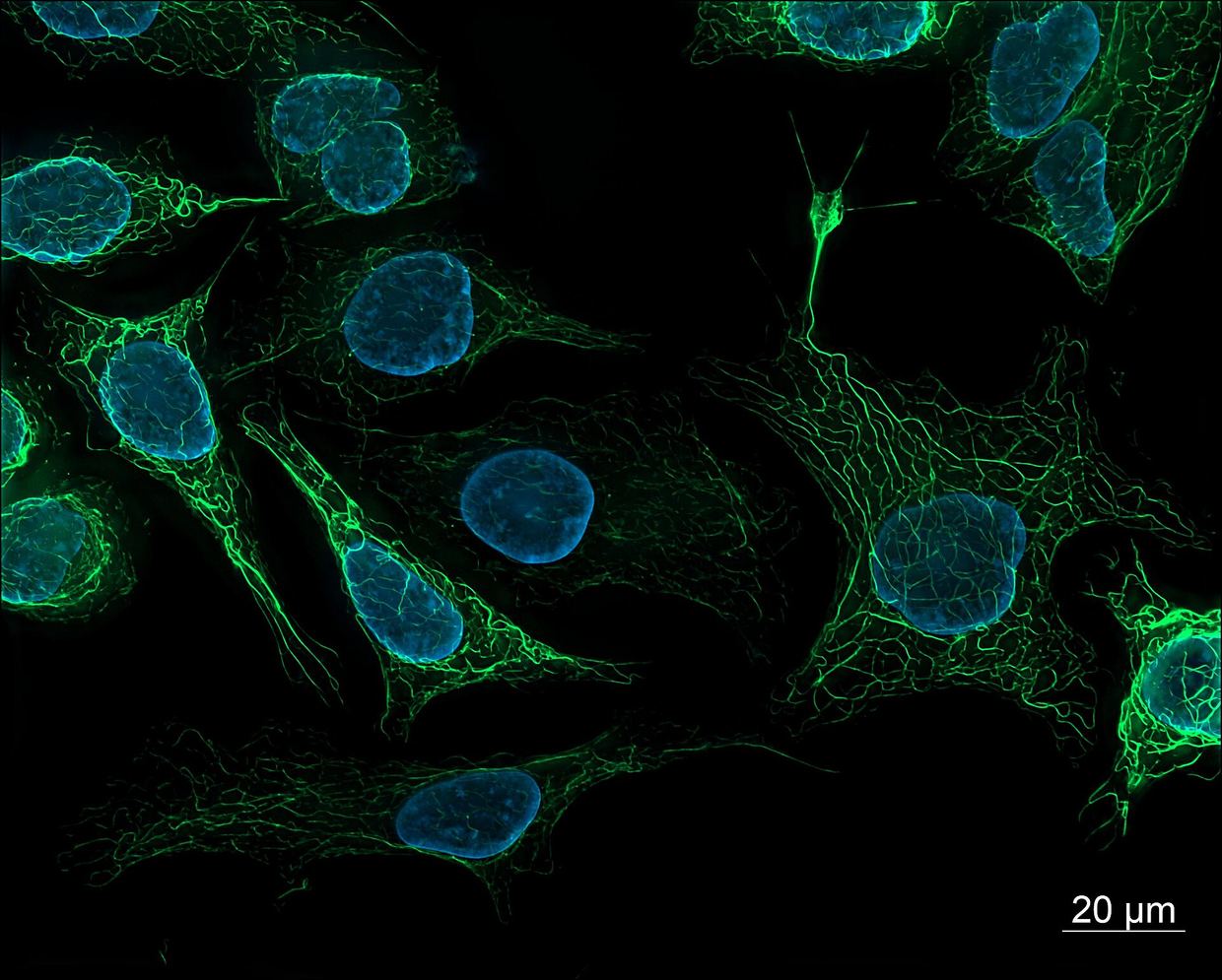
The FLIM-SIM method images fine cellular structures and reports their molecular environment, such as pH. It was demonstrated on a human bone cancer cell line.
Researchers from Bielefeld University and the University of Vienna have developed a microscopy technique that makes the smallest cellular structures visible and at the same time shows how their molecular environment changes, news-medical.net reports. The method, called FLIM-SIM, was created within the European CHEM-SCAN project, and the results were published in the journal Optica. news-medical.net
news-medical.netnews-medical.netOpen source ↗ According to the report, it reaches a spatial resolution of 156 nanometers and can capture rapid processes in living cells.
news-medical.net
news-medical.netnews-medical.netOpen source ↗
How FLIM-SIM combines two techniques
The approach joins two methods. Structured illumination microscopy (SIM) makes visible cellular structures that are too small to be resolved by conventional light microscopes. Fluorescence lifetime imaging microscopy (FLIM) measures how long fluorescent molecules emit light, and this lifetime can change with the molecular environment, for example when pH changes. news-medical.net
news-medical.netnews-medical.netOpen source ↗
Until now, this information could be combined with high-resolution microscopy only to a limited extent. The new technique offers high spatial resolution, fast imaging rates and a large field of view. The researchers demonstrated these capabilities on a human bone cancer cell line. news-medical.net
news-medical.netnews-medical.netOpen source ↗
Dr. Henning Ortkraß, one of the study's authors, said that super-resolution combined with lifetime measurements shows not only where certain molecules are located in a cell, but also the molecular environment they are in. news-medical.net
news-medical.netnews-medical.netOpen source ↗
Use in drug research
The method is being applied in CHEM-SCAN, which investigates, among other things, how chemotherapeutic agents used in cancer treatment can be adapted to act as effectively as possible on both tumours and the metastases that arise from them. To study efficacy, the project uses specialised fluorescent dyes that, with FLIM measurements, give information on the state of cancer cells. news-medical.net
news-medical.netnews-medical.netOpen source ↗
The authors note that the better researchers can observe what drugs trigger inside individual cells, the more precisely they can study the effects and potential harmful effects of those drugs. They suggest the technique could help improve understanding of drug action at the cellular level. news-medical.net
news-medical.netnews-medical.netOpen source ↗
Professor Thomas Huser, who coordinates CHEM-SCAN and leads the research group, said the results open a new dimension of contrast for super-resolution microscopy: fine cell structure can be studied at high speed together with information on the molecular environment. In his words, this is an important basis for future live-cell and drug research. news-medical.net
news-medical.netnews-medical.netOpen source ↗