Stanford bioengineers control actomyosin contractility using blue light
Engineered filamentous myosins change velocity and modulate cellular contraction under blue light in vitro and in live cells.
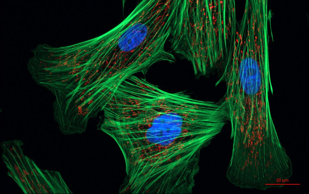
Bioengineers from Stanford University have developed engineered filamentous myosins that alter their speed, direction, and interaction with actin in response to blue light, according to a study published in Nature Communications doi.org
Nature Communicationsdoi.orgOpen source ↗. The system expands the MyLOV family of genetically engineered motors to create functional minifilaments capable of precise optical control of actomyosin contractility
doi.org
Nature Communicationsdoi.orgOpen source ↗.
In vitro speed and contraction control
To evaluate the motor dynamics, the researchers performed in vitro single-molecule tracking assays and reconstituted actin network contractility experiments doi.org
Nature Communicationsdoi.orgOpen source ↗. The gearshifting minifilaments changed velocity and direction under blue light illumination
doi.org
Nature Communicationsdoi.orgOpen source ↗. When embedded in reconstituted actin networks, these engineered proteins directly modulated network contraction rates based on optical input
doi.org
Nature Communicationsdoi.orgOpen source ↗. The team also created an alternative minifilament design that interacts processively with actin exclusively when exposed to blue light
doi.org
Nature Communicationsdoi.orgOpen source ↗.
Optogenetic regulation in live cells
The authors further characterized the contractile phenotypes of the engineered minifilaments in live cells doi.org
Nature Communicationsdoi.orgOpen source ↗. Optical stimulation enabled targeted control of protrusion retraction, cortical flow, and whole-cell migration
doi.org
Nature Communicationsdoi.orgOpen source ↗. The engineered filamentous myosins provide an optogenetic toolset to examine self-organization, force transmission, and mechanotransduction in dynamic contractile systems across cellular and tissue models
doi.org
Nature Communicationsdoi.orgOpen source ↗.