NoRI microscopy quantified protein and lipid changes in mouse kidneys
A label-free stimulated Raman imaging technique mapped kidney biomass at high resolution in mouse models without chemical staining.
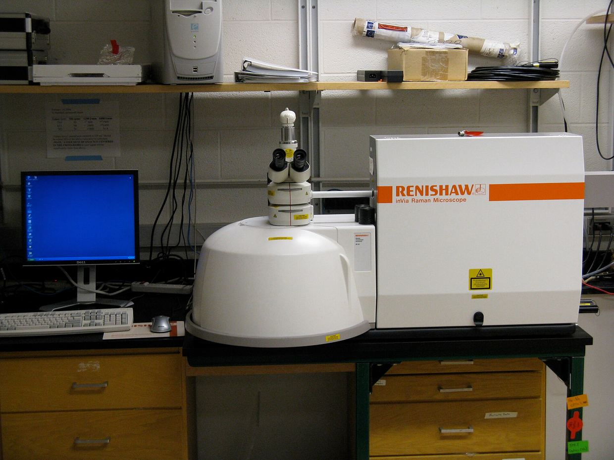
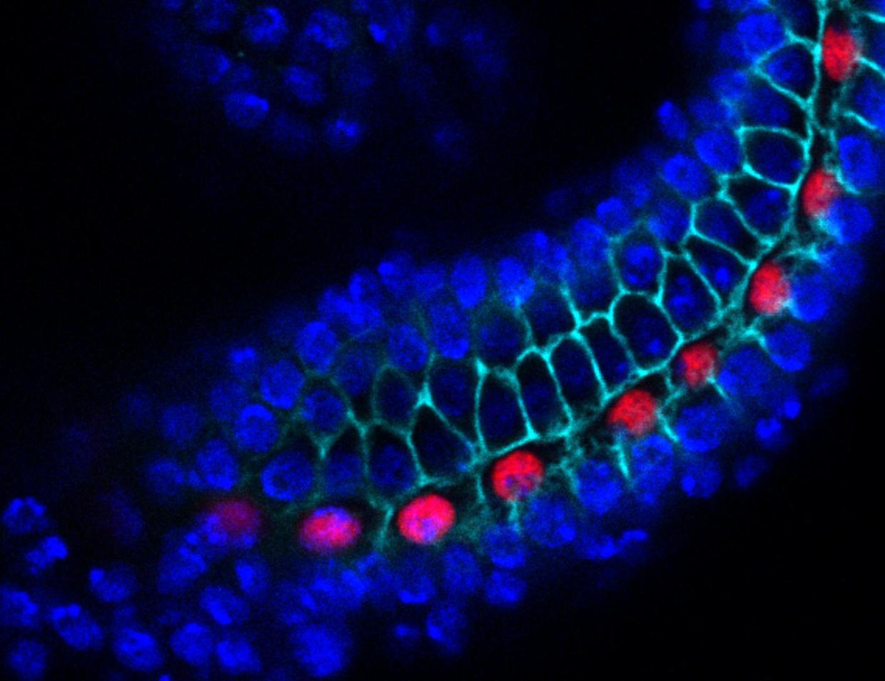
Researchers have demonstrated Normalized Stimulated Raman Imaging (NoRI) to quantitatively measure protein, lipid, and water content in tissue sections at high spatial resolution without chemical staining, according to a study published in the Proceedings of the National Academy of Sciences doi.org
Proceedings of the National Academy of Sciencesdoi.orgOpen source ↗. Standard histopathological preparation techniques often distort native cellular architecture and wash away lipids, limiting reproducibility in quantitative diagnostic analyses.
In experiments on mouse kidney sections, the platform preserved tissue architecture and computationally corrected Raman signals to quantify absolute biomass concentrations doi.org
Proceedings of the National Academy of Sciencesdoi.orgOpen source ↗. The acquired imaging data allowed classification of tubule types with an F1 score of 0.93, anatomical regions with an F1 score of 0.91, and biological sex with an F1 score of 0.97
doi.org
Proceedings of the National Academy of Sciencesdoi.orgOpen source ↗. The measurements revealed that female mouse tubules contained 6.9 mg/mL more cytoplasmic lipids (p=0.028) and 26.3 mg/mL more nuclear proteins (p<0.001) compared to male samples.
The system also tracked tissue changes over 25 days in mouse models of acute kidney injury doi.org
Proceedings of the National Academy of Sciencesdoi.orgOpen source ↗. It quantified brush border remodeling alongside lipid droplet dynamics, achieving an F1 score of 1.0 for feature classification based on spatial lipid distribution
doi.org
Proceedings of the National Academy of Sciencesdoi.orgOpen source ↗. The authors reported that the approach provides a reproducible platform for pathology workflows and tissue feature discovery.