BrainSPACE pipeline reveals layer 4 neuronal diversity in macaque visual cortex
Researchers mapped roughly 100,000 nuclei across visual areas in rhesus macaques, uncovering area-specific principal neuron subtypes in layer 4.
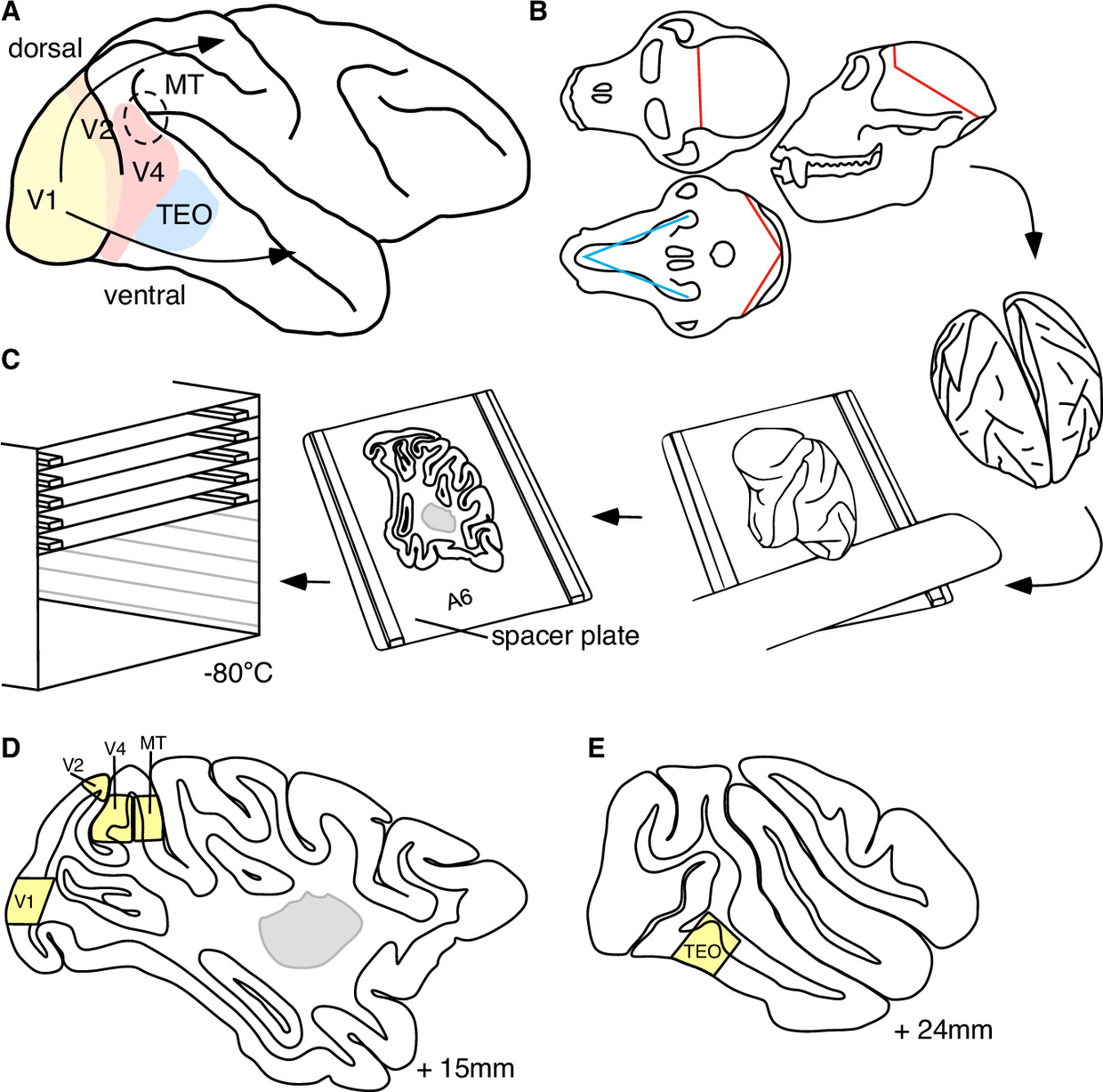
Neuroscientists have developed a tissue-banking workflow called BrainSPACE to map the visual cortex of the rhesus macaque at single-nucleus resolution, as reported in Nature Communications. Applying single-nucleus RNA sequencing (snRNA-seq) to approximately 100,000 nuclei from visual areas V1 and V4, the team established a comparative transcriptomic atlas of early and mid-level visual processing regions doi.org
Nature Communicationsdoi.orgOpen source ↗.
Tissue archiving and profiling
To preserve RNA integrity for sequencing and imaging, whole brains were extracted within 30 minutes of cardiac arrest and placed in ice-cold buffer. Hemispheres were separated along the midline and sectioned into 3 mm sagittal slices on precooled spacer plates. These slices were imaged, aligned to an anatomical reference atlas, and stored at -80 °C in custom BrainSPACE containers. The researchers isolated 5 to 10 by 10 mm tissue blocks spanning all cortical layers and underlying white matter across five visual regions: primary visual cortex (V1), visual area 2 (V2), visual area 4 (V4), middle temporal area (MT), and area TEO doi.org
Nature Communicationsdoi.orgOpen source ↗.
Divergence in layer 4 principal neurons
Transcriptomic profiling revealed that while inhibitory GABAergic interneurons remained largely conserved across visual regions, excitatory principal neurons displayed pronounced area-specific divergence doi.org
Nature Communicationsdoi.orgOpen source ↗. The snRNA-seq dataset identified seven distinct principal neuron subtypes localized specifically to layer 4 of V1, compared to a single layer 4 subtype unique to V4
doi.org
Nature Communicationsdoi.orgOpen source ↗. Single-molecule fluorescent in situ hybridization (smFISH) across all five sampled areas verified that marker genes followed spatial expression gradients that match the hierarchical structure of both ventral and dorsal visual processing streams
doi.org
Nature Communicationsdoi.orgOpen source ↗.
Gene ontology evaluations showed enrichment of synaptic plasticity pathways among the unique layer 4 principal neurons doi.org
Nature Communicationsdoi.orgOpen source ↗. The analysis highlighted differential expression of neurodevelopmental disorder risk genes, such as NTNG1 and NLGN1, linking molecular divergence in layer 4 circuitry to visual pathway specialization in primates
doi.org
Nature Communicationsdoi.orgOpen source ↗.