SORDINO fMRI method acquires silent, distortion-free brain images in awake mice at 9.4 T
The zero echo time method, tested at 9.4 T, keeps scanner noise near idle level and allows brain-wide imaging of awake, behaving mice.
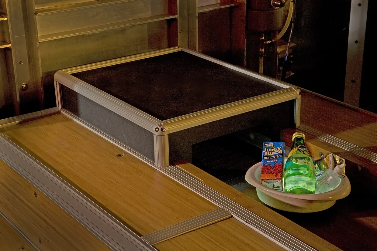
Researchers have described SORDINO, a zero echo time (ZTE) based functional MRI method that works silently and without distortion, in Nature Neuroscience. At 9.4 T the authors compared it with conventional gradient-recalled-echo echo-planar imaging (EPI) and standard ZTE, and then used it for brain-wide imaging in awake, behaving mice nature.com
Nature Neurosciencenature.comOpen source ↗. The name stands for Steady-state On-the-Ramp Detection of INduction-decay with Oversampling.
After an initial ramp, the method keeps the gradient amplitude nearly constant while its direction rotates continuously, and data are acquired only as the direction changes. Excitation uses nonselective radiofrequency pulses with a phase increment of 117 degrees, which serves as radiofrequency spoiling.
Acoustic noise, distortion and recording compatibility
In rat imaging the average slew rate was 0.21 T/m/s, versus 15.66 T/m/s for standard ZTE and 1,263.62 T/m/s for EPI at the same spatiotemporal resolution. Sound pressure measurements up to 100 kilohertz in a 9.4 T scanner showed SORDINO at levels practically indistinguishable from an idle scanner, while EPI and standard ZTE produced pronounced acoustic peaks. In phantoms, SORDINO showed no ghosting and negligible spatial displacement against a turbo spin echo reference, whereas EPI was visibly warped. Lower receiver bandwidths gave up to an eightfold gain in temporal signal-to-noise ratio over standard ZTE.
Electromagnetic interference was also low. Simulated neuronal spikes generated inside the magnet could be sorted online in real time, and fast-scan cyclic voltammetry with a dummy cell showed stable background currents. A miniature calcium microscope over the prelimbic cortex of an awake mouse recorded single-cell activity during scanning, and event amplitudes and baselines were preserved between idle and active periods nature.com
Nature Neurosciencenature.comOpen source ↗.
Contrast mechanism and awake mice
In freshly drawn blood, deoxygenated venous blood gave a higher SORDINO signal than oxygenated arterial blood, the reverse of what EPI shows. The authors report that the contrast reflects changes in longitudinal relaxation tied to cerebral blood volume, tissue oxygenation and blood inflow, rather than BOLD dephasing. Gas challenge experiments with fiber photometry and voltammetry showed that the signal follows tissue oxygen and blood volume.
With head plates that also served as radiofrequency transceivers, mice were habituated to head fixation within five days. Serum cortisol decreased significantly by day three and matched non-imaged controls by day five, while mice scanned with EPI showed sustained elevations and lost body weight. Resting-state imaging in 25 awake mice showed low framewise displacement and recovered the default mode, salience and lateral cortical networks, consistent with Allen Mouse Brain Atlas tract-tracing. In a reach-and-grasp task with head-fixed mice, activation began in the contralateral primary motor cortex together with deactivation in the retrosplenial cortex.
Sources · 2
Found a mistake?Read also