Enzyme-free laser microdissection (Leica LMD7) profiles proteins of α-, β- and δ-cells in mouse islets
Authors isolated islet cells from fresh-frozen mouse pancreas without enzymes or fixation. They report that enzymatic digestion alters expression of membrane-associated proteins.
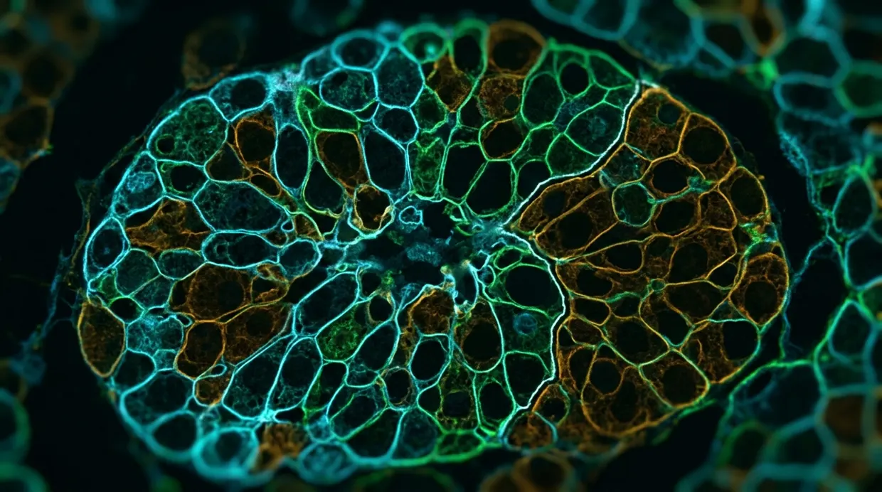
A nonenzymatic method for isolating pancreatic islet cells by laser microdissection (Leica LMD7) is described in Experimental & Molecular Medicine. It was used to collect α-, β- and δ-cells from fresh-frozen, unfixed mouse pancreatic tissue for proteomic and transcriptomic analysis nature.com
Experimental & Molecular Medicinenature.comOpen source ↗. According to the authors, the approach avoids enzymatic digestion and chemical fixation and preserves the native molecular state of the cells.
Why enzymatic isolation changes the data
Standard workflows dissociate tissue with trypsin and sort the cells by fluorescence-activated cell sorting. The study compared its results with such data compiled by other groups. The authors report that enzymatic digestion alters gene expression patterns, particularly those of membrane-associated proteins.
The team also dropped paraformaldehyde fixation, which left RNA fragmented and prevented mass spectrometric detection of proteins. Tissue for proteomics was embedded in a sucrose medium instead of the standard optimal cutting temperature compound, because the polymers in that compound interfere with liquid chromatography-tandem mass spectrometry. Cells were identified by a rapid immunofluorescence staining at room temperature, and Swiss Webster mice were chosen for their larger islets.
What the cell profiles showed
For proteomics, approximately 4,100 α-cells, 20,000 β-cells and 600 δ-cells were collected. The analysis identified 660 proteins in α-cells, 1,335 in β-cells and 1,765 in δ-cells. Of these, 13 were unique to α-cells, 247 to β-cells and 795 to δ-cells nature.com
Experimental & Molecular Medicinenature.comOpen source ↗.
For transcriptomics, approximately 4,800 α-cells, 8,400 β-cells and 600 δ-cells were collected. The analysis identified 18,394 transcripts in α-cells, 11,696 in β-cells and 18,071 in δ-cells.
The authors identified cell-type-specific proteins that are typically underrepresented in pancreatic single-cell transcriptomic datasets. β-cells were enriched in vesicle trafficking proteins, α-cells showed distinct calcium-dependent action potential machinery, and δ-cells showed elevated expression of focal adhesion-related proteins. They also report an inverse molecular relationship between β- and δ-cells, potentially driven by transcriptional regulators such as Mlxipl.
The authors present these profiles from intact tissue as a reference point for future comparisons in diabetes and other endocrine disorders nature.com
Experimental & Molecular Medicinenature.comOpen source ↗.