Astrocytes in mouse cell culture truncate α-synuclein with cathepsin D and enlarge aggregates in neurons
In a mouse neuron–astrocyte co-culture, astrocytes cleaved internalized α-synuclein fibrils and passed material to neurons. Pepstatin A reduced the larger aggregates.
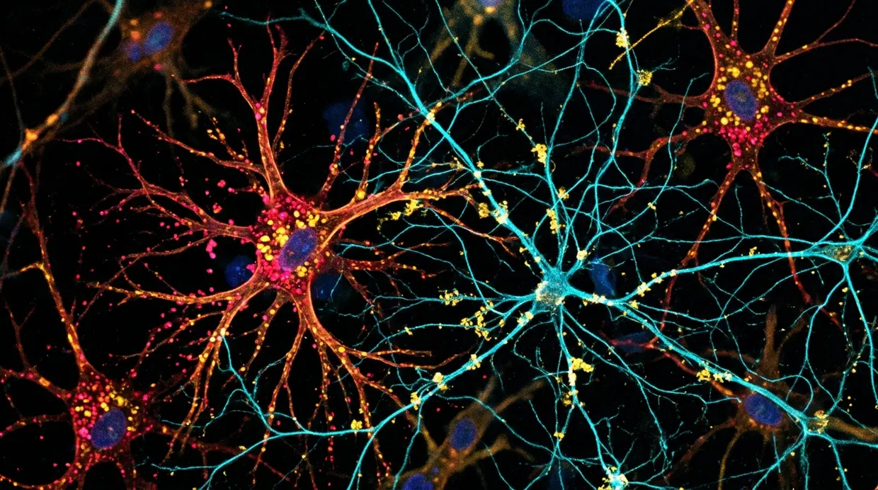
Researchers reported in EMBO Reports that astrocytes in a mouse neuron–astrocyte co-culture process α-synuclein pre-formed fibrils (PFFs) and influence how aggregates in neurons mature nature.com
EMBO Reportsnature.comOpen source ↗. The model combined primary mouse hippocampal neurons with neurosphere-derived astrocytes and reproduced the contacts of a tripartite synapse, according to the authors. Astrocytes were added to established neuronal networks at day in vitro 10, and cultures were kept until day 24.
Parkinson's disease and related synucleinopathies are marked by accumulation and spread of α-synuclein aggregates, a process studied mostly in neurons. Whether astrocytes actively take part in it had remained unclear.
Astrocytes truncate and pass on α-synuclein
Astrocytes internalized PFFs and generated C-terminally truncated α-synuclein species through a process dependent on cathepsin D (CtsD). With an antibody to residues 91–99, full-length protein and lower-molecular-weight fragments were detected as early as 6 hours after treatment. An antibody to residues 118–123 did not recognize the fragments, and the full-length protein declined as they accumulated. In neuron-only cultures, truncated fragments appeared only in the insoluble fraction.
PFF exposure caused lysosomal membrane damage, a senescence-like reactive state and CtsD upregulation in astrocytes. PFF-containing lysosomes were engulfed by autophagy but persisted, and a subset was found beyond the astrocytic membrane boundary nature.com
EMBO Reportsnature.comOpen source ↗.
Astrocytes preloaded with 0.5 µg/mL PFFs for 24 hours and then co-cultured with naïve neurons led to pS129-α-synuclein-positive aggregates along neurites after 14 days. Neurons with untreated astrocytes showed no such signal. Labeled fibril puncta were found along axons, partly next to VGLUT1-positive presynaptic and HOMER1-positive postsynaptic markers.
Effect on aggregate size
When neurons were challenged with 0.5 µg/mL PFFs and analyzed at 6 or 10 days, co-cultures had fewer punctate aggregates at day 6, but the numbers were comparable by day 10. By day 10, Lewy neurite-like structures were markedly longer and larger in co-cultures, while puncta size and total aggregate counts did not increase.
Pepstatin A, an aspartic protease inhibitor, reduced truncated fragments in astrocytes in a dose-dependent way at 20 and 100 µM. In co-cultures, 10 µM pepstatin A reduced Lewy neurite-like aggregates to the size seen in neuron-only cultures, without affecting puncta. In neuron-only cultures it produced no significant change nature.com
EMBO Reportsnature.comOpen source ↗.
The authors state that their findings support a model in which astrocytic lysosomal remodeling of internalized α-synuclein contributes to maturation and spread of neuronal pathology in this co-culture system.