In mice, Rickettsia parkeri passes from an Amblyomma maculatum tick within 1 hour of attachment
In a single-tick mouse model, the bacterium spread to lymph nodes and internal tissues early, while the skin's pro-inflammatory response appeared by 144 hours.
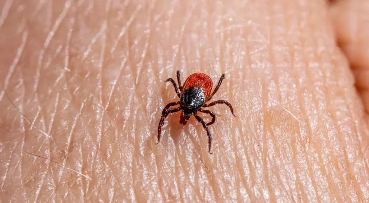
Researchers Chanakan Suwanbongkot and Kevin Macaluso at the University of South Alabama studied how Rickettsia parkeri, an emerging tick-borne pathogen, is transmitted by Amblyomma maculatum, and reported the results in PLoS Pathogens. In mice, the bacterium passed from an infected tick to the host within 1 hour after attachment and spread rapidly to lymph nodes and internal tissues plos.org
PLoS Pathogensplos.orgOpen source ↗.
Transmission model and bacterial load
Earlier work on rickettsial pathogenesis in mice mostly relied on needle inoculation. As a result, transmission dynamics and the influence of tick saliva on the host response at the infection site were undefined. During feeding, tick salivary factors manipulate the host's hemostatic and immune responses.
To address this, the authors developed a single-tick feeding model. It tracked transmission and dissemination from the early stage of attachment, 1 hour, to the late feeding stage, 144 hours.
The quantifiable bacterial load in mouse skin delivered by an infected tick was 9,500 bacteria at 3 hours. It increased to 15,000 at 48 hours and to 22,000 rickettsiae at 144 hours plos.org
PLoS Pathogensplos.orgOpen source ↗.
Cutaneous immune response
The team characterized the skin response by immunophenotyping, cytokine and chemokine profiling, and histopathological analysis. The working hypothesis was that the host response would differ depending on whether the tick was infected.
At 3 and 48 hours after attachment, the degree of time-dependent inflammation was similar regardless of the tick's infection status.
At 144 hours the picture differed. In skin infected with R. parkeri, macrophages were the dominant cell type, levels of IFN-γ and TNF-α were higher, and production of IL-10 was reduced plos.org
PLoS Pathogensplos.orgOpen source ↗.
The authors interpret this as delayed pro-inflammatory activation in response to rickettsial infection delivered by ticks. They state that the study gives insight into the early events of Rickettsia transmission and the host response, and provides a basis for developing preventive strategies against tick-borne rickettsial disease. All data come from a mouse model.