Tulane University researchers describe previously undocumented muscle attachment near the hip
Graduate students identified a previously undescribed anatomical variation above the hip joint, confirmed via histological analysis in human cadavers.
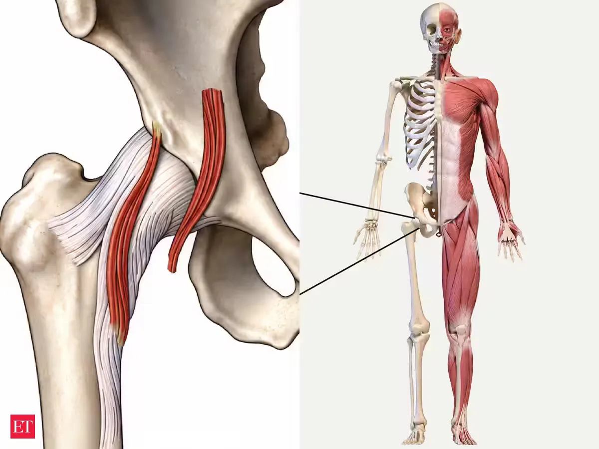
Graduate students Dominic Roy and Cooper Bruce, working under Professor R. Shane Tubbs at Tulane University, identified an undocumented muscle attachment situated above the human hip joint during cadaver dissections wwno.org
wwno.orgwwno.orgOpen source ↗. The discovery, reported by wwno.org, emerged while dissecting deep pelvic structures in the university's anatomy laboratory.
After finding the structure absent from anatomical literature, the team examined hundreds of cadaver sides across Tulane laboratories. International collaborators in South Korea, Japan, and Europe corroborated the findings by identifying the identical muscular variant in their own anatomical dissection cohorts.
Morphological and Histological Characteristics
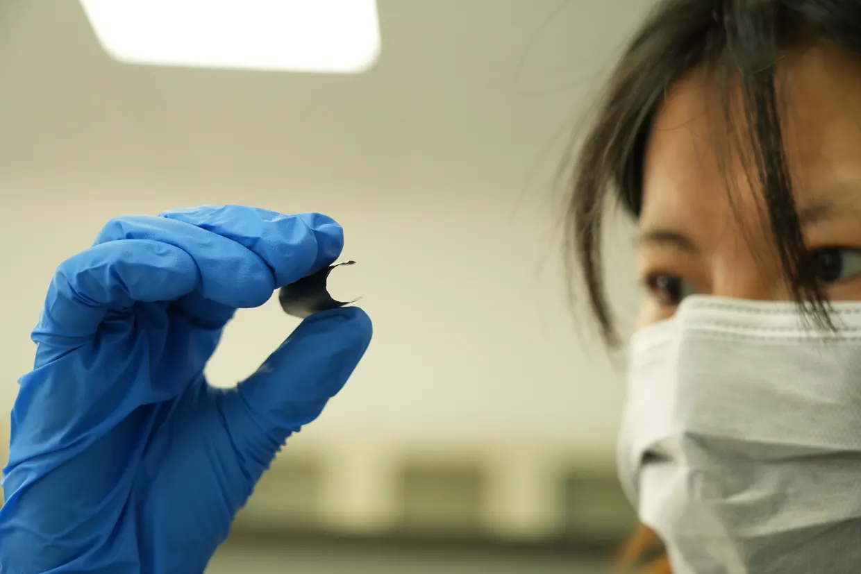
The identified structure is located near the inguinal region of the pelvis and exhibits a close physical relationship with the anterior capsule of the hip joint. To exclude pathological tissue changes or non-muscular anomalies, the researchers prepared histological cross-sections of the specimens.
Microscopic examination confirmed that the structure consists of normal skeletal muscle tissue rather than fibrous or pathological alterations wwno.org
wwno.orgwwno.orgOpen source ↗. Histological findings further verified its direct attachment to the surrounding hip joint capsule. The variation does not appear in every examined donor, which the authors noted is standard for non-pathological anatomical variants.
Potential Clinical Implications and Next Steps
The team noted similarities between this structure and adjacent muscular layers known to cause anterior hip pain independent of joint pathology. The authors suggest that this newly documented attachment might contribute to regional pain syndromes or conditions like snapping hip syndrome, though clinical verification is currently lacking.
Professor Tubbs emphasized that research remains in its initial morphological characterization phase. Planned next steps involve testing whether the muscular slip can be identified through diagnostic magnetic resonance imaging (MRI). Subsequent investigations aim to evaluate symptomatic patients to correlate clinical presentations with the presence of this anatomical variation.