[18F]FET PET shows 91.2% sensitivity for glioblastoma vs 52.6% for contrast MRI in 61 patients
In 472 histology-verified tissue samples from 61 patients with IDH-wildtype glioblastoma, amino acid PET found tumor that contrast-enhanced MRI missed.
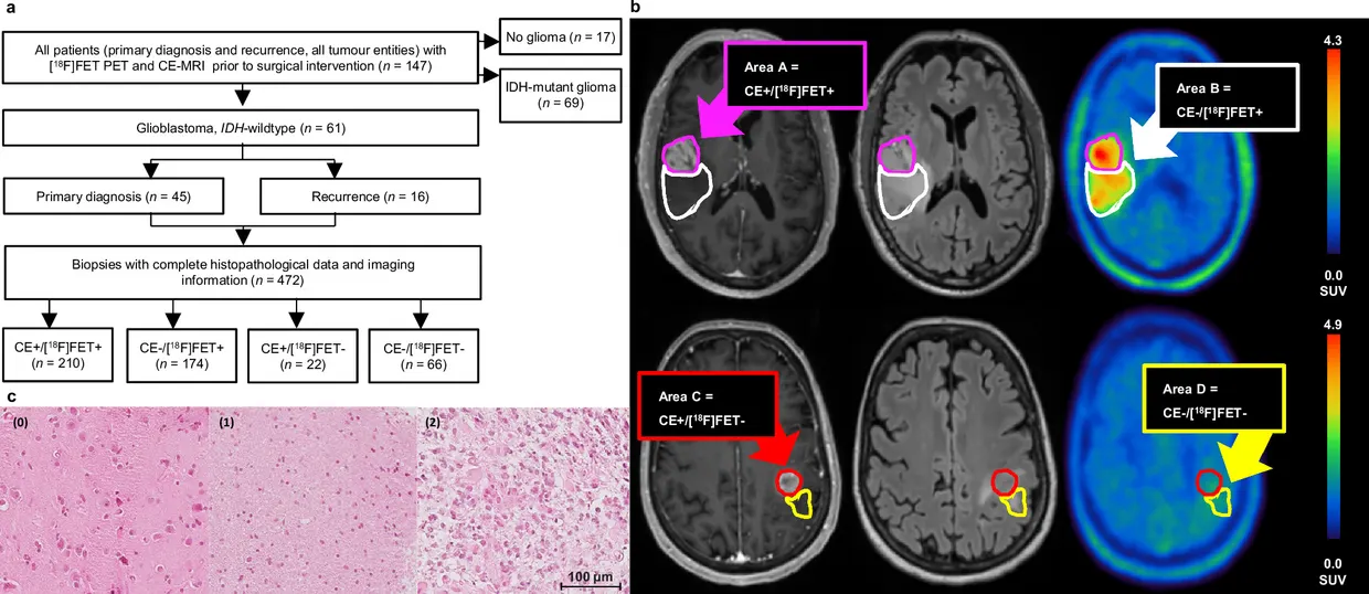
A study in Nature Communications compared O-(2-[18F]fluoroethyl)-L-tyrosine PET ([18F]FET PET) with contrast-enhanced MRI (CE-MRI) in 61 patients with IDH-wildtype glioblastoma (WHO grade 4, 2021 classification). The analysis used 472 spatially matched tissue samples from stereotactic biopsies and resections, each verified by histology. nature.com
Nature Communicationsnature.comOpen source ↗
According to the authors, [18F]FET PET detected tumor tissue significantly better than CE-MRI, especially in regions without enhancement on MRI. Sensitivity for tumor detection was 91.2% for PET versus 52.6% for CE-MRI.
Cohort and sampling
All patients had PET and CE-MRI before surgery. Of the 61 patients, 45 (73.8%) had newly diagnosed disease and 16 (26.2%) had recurrence. Tissue was obtained by stereotactic biopsy in 26 patients (42.6%) and together with partial or gross total resection in 35 (57.4%). Median age was 59 years (range 33–84), and the median interval between PET and MRI was 1 day (range 0–28). A subgroup limited to an interval of up to 14 days gave results concordant with the main analysis.
The median was 5 samples per patient (range 1–27). Quantitative imaging data were missing for 8 biopsies because of intraoperative technical issues. Samples were assigned to four areas by PET positivity (tumor-to-background ratio of at least 1.6) and by contrast enhancement: 210 samples (44.5%) were enhancing and PET-positive, 174 (36.9%) non-enhancing and PET-positive, 22 (4.6%) enhancing and PET-negative, and 66 (14.0%) negative on both.
Detection beyond contrast enhancement
PET uptake was present in all 61 patients (100%), while contrast enhancement was seen in 56 of 61 (91.8%). In every enhancing case, tracer uptake extended beyond the enhancing region. nature.com
Nature Communicationsnature.comOpen source ↗
In non-enhancing tissue, PET-positive samples had a positive predictive value of 98.3% for histologically confirmed tumor. The authors report that the results were consistent in newly diagnosed and recurrent disease and in both the resection and biopsy groups.
The authors conclude that [18F]FET PET complements MRI by revealing tumor burden not seen on MRI, and suggest that adding amino acid PET to routine planning may improve the accuracy of tumor visualization.
Sources · 1
Found a mistake?Read also