Single-cell study of fat tissue in 58 people with obesity compares those with and without type 2 diabetes
Researchers profiled paired omental and subcutaneous adipose samples from people with obesity, with and without T2D, and found depot-specific changes in stromal and immune cells.
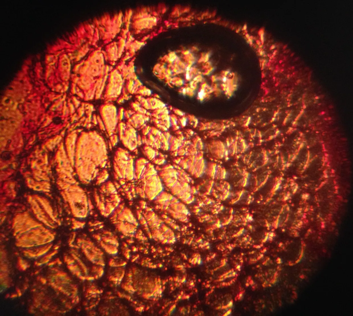
A study in Science Advances examined 58 individuals with obesity, with or without a clinical diagnosis of type II diabetes (T2D). The authors profiled paired samples of omental and subcutaneous adipose tissue by single-cell RNA sequencing (n = 10), flow cytometry under basal and stimulated conditions (n = 42), and histology (n = 17) science.org
Science Advancesscience.orgOpen source ↗. Flow cytometry and chemokine quantification were also performed in paired whole blood.
The authors identified reprogramming of the omental stromal compartment, centered on mesothelial and endothelial cells, alongside contrasting innate T cell and macrophage phenotypes across the two depots. Mesothelial cells and IGFBP2+ cells showed a fibrotic, epithelial-mesenchymal transition–like phenotype. In T2D, omental endothelial cells showed a lipid-overload, inflammatory, antigen presentation profile.
Innate T cells in omental and subcutaneous adipose tissue had divergent inflammatory profiles between the tissues. According to the authors, the findings give previously unidentified insights into the pathophysiology of T2D in adipose tissue and identify compensatory networks within the tissues that could be exploited by targeted therapeutics.