Chiba University splits 8×8 mm CVD diamond along {100} plane with 10 ps laser pulses
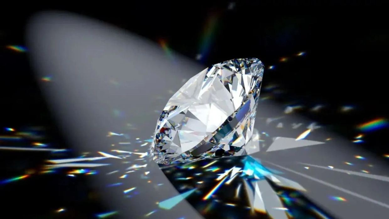
In a laboratory test, Chiba researchers split one 8 mm × 8 mm CVD diamond along the {100} plane after four steps of 10 ps laser pulses.
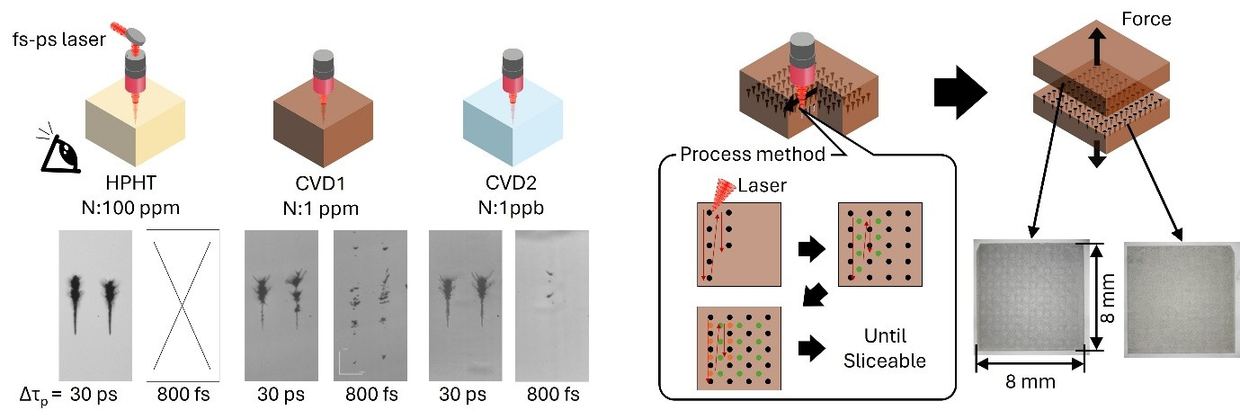
A team led by Professor Hirofumi Hidai at the Graduate School of Engineering, Chiba University (Japan), studied how lab-grown single-crystal diamonds respond to ultrashort laser pulses. Based on the results, they split an 8 mm × 8 mm CVD diamond along the {100} plane, according to cn.chiba-u.jp cn.chiba-u.jp
cn.chiba-u.jpcn.chiba-u.jpOpen source ↗. The paper, co-authored by Mahiro Ikeno, appeared in Diamond and Related Materials, volume 169, on November 1, 2026
cn.chiba-u.jp
cn.chiba-u.jpcn.chiba-u.jpOpen source ↗.
How HPHT and CVD diamonds respond to the laser
The researchers compared three single-crystal diamonds: one grown by the high-pressure high-temperature (HPHT) method and two by chemical vapor deposition (CVD). The samples differed in nitrogen impurity content. Focused pulses with durations from 233 fs to 30 ps were applied, and the modified regions inside the crystals were examined by optical microscopy, spectroscopy and image analysis cn.chiba-u.jp
cn.chiba-u.jpcn.chiba-u.jpOpen source ↗. In the reported setup, the pulse energy was 2 µJ, with ten pulses delivered to each spot.
The samples behaved differently. The two CVD diamonds could be modified with much shorter pulses than the HPHT diamond cn.chiba-u.jp
cn.chiba-u.jpcn.chiba-u.jpOpen source ↗. In the HPHT crystal, no internal changes were visible at durations below 7 ps. In the CVD crystals, modification began at 233 fs in the first and at 800 fs in the second.
In the CVD diamonds, the shortest pulses produced tiny, separated marks, while pulses of about 10 ps or longer produced more continuous modified regions cn.chiba-u.jp
cn.chiba-u.jpcn.chiba-u.jpOpen source ↗. Nitrogen content alone did not explain the differences. The authors suggest that microscopic defects linked to each crystal's growth history may play a role. CVD diamonds also tended to show less stable modified regions and greater variation in their shape than HPHT diamonds
cn.chiba-u.jp
cn.chiba-u.jpcn.chiba-u.jpOpen source ↗.
Slicing in four steps
For slicing, the team applied 10 ps pulses to an 8 mm × 8 mm CVD diamond and formed modified regions in a controlled grid. Instead of trying to create a full crack network at once, they added new modified regions at the midpoints between existing ones cn.chiba-u.jp
cn.chiba-u.jpcn.chiba-u.jpOpen source ↗. After four such steps, the crystal split cleanly along the {100} plane under a light tap from tweezers. This plane is the easiest to polish, which matters for practical use
cn.chiba-u.jp
cn.chiba-u.jpcn.chiba-u.jpOpen source ↗.
The area of the resulting slice was approximately 710 times larger than that of the 300 µm-square HPHT diamond slice obtained in the group's earlier study cn.chiba-u.jp
cn.chiba-u.jpcn.chiba-u.jpOpen source ↗. The authors regard this as a step toward the larger diamond substrates needed for optical components and electronics. So far, the method has been demonstrated on one such crystal.
Professor Hidai named possible applications: substrates and components for high-power laser optical windows in lithography systems, heat spreaders for advanced electronics, and diamond-based power semiconductors cn.chiba-u.jp
cn.chiba-u.jpcn.chiba-u.jpOpen source ↗. The researchers note that the slicing steps are potentially scalable and envision adapting the method for industrial use
cn.chiba-u.jp
cn.chiba-u.jpcn.chiba-u.jpOpen source ↗.