Scientists grow human lens organoids from stem cells in vitro
Researchers modeled human lens development in vitro using induced pluripotent stem cells, creating mini lenses with authentic tissue architecture.
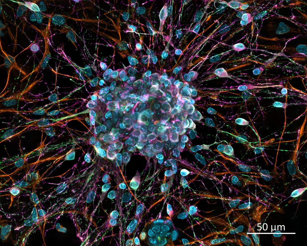
Researchers from Anglia Ruskin University, Osaka University, and Cardiff University have developed an in vitro platform to grow miniature human lens structures from human induced pluripotent stem cells (hiPSCs). According to the study published in npj Regenerative Medicine, the team combined spatially patterned self-formed ectodermal autonomous multizone (SEAM) differentiation with a specialized lens optimized medium (LOM) doi.org
npj Regenerative Medicinedoi.orgOpen source ↗.
The resulting lentoids exhibited zone-specific structural organization, capsule basement membrane deposition, and upregulation of key lens crystallins and connexins. Ultrastructural imaging revealed classic hallmarks of a maturing natural lens, including elongated cortical-like fibers, organized epithelial cell layers, and cadherin-mediated intercellular junctions featuring ball-and-socket-like interdigitations doi.org
npj Regenerative Medicinedoi.orgOpen source ↗.
Cellular Plasticity and Secondary Lentoids
The authors noted that the system sustained self-renewing, preplacodal, and presumptive lens ectodermal cell populations expressing PAX6, SIX3, and SOX2. Following the activation of differentiation transcription factors PROX1 and SOX1, these progenitor-like populations formed secondary lentoids in vitro doi.org
npj Regenerative Medicinedoi.orgOpen source ↗.
Platform for Disease Modeling
Because direct models of human lens organogenesis have remained limited, researchers plan to apply this platform to explore cataractogenesis, protein homeostasis, and cellular differentiation pathways. Cataracts, which occur when structural lens proteins clump together and scatter light, currently lack pharmacological interventions and are managed exclusively by surgical removal.