Adaptive optics OCT shows 4.16–6.86% yearly retinal ganglion cell loss in glaucoma
In a preliminary 3–5 year study, AO-OCT imaged single retinal ganglion cells and flagged glaucoma progression earlier than standard OCT and visual field tests.
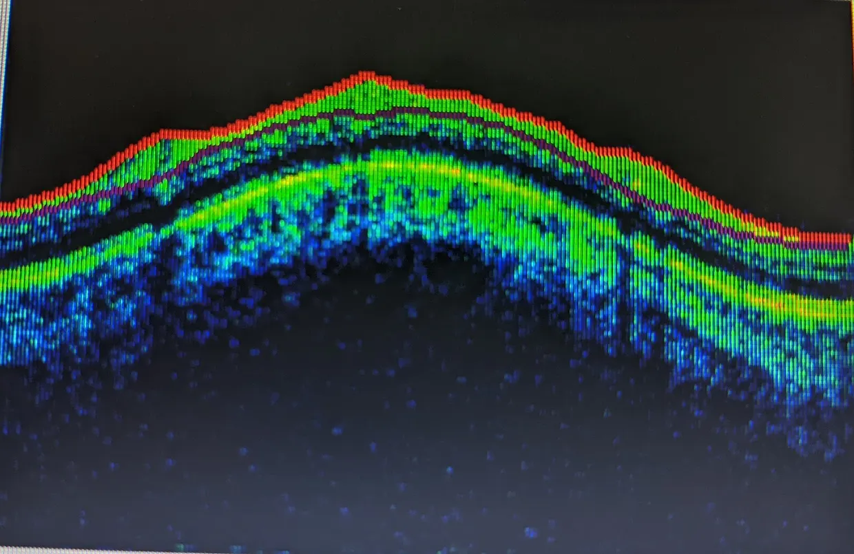
A preliminary study in Science Advances used high-resolution adaptive optics–optical coherence tomography (AO-OCT) to track individual retinal ganglion cells in glaucoma patients and healthy volunteers over 3 to 5 years science.org
Science Advancesscience.orgOpen source ↗. Glaucoma is a leading cause of irreversible blindness and involves progressive loss of these cells. The authors note that current clinical methods often identify the disease only after substantial damage has occurred.
In glaucoma, cell density decreased by 4.16 to 6.86% per year, versus 0.27% per year in the healthy volunteer group. Neurodegeneration in glaucoma was higher than in normal aging.
AO-OCT detected progression 0.74 years before conventional OCT and 1.49 years before visual field testing. The authors describe it as a more sensitive tool for monitoring glaucomatous damage with higher precision. The work is preliminary.
出典 · 1
誤りを見つけましたか?あわせて読みたい