Manganese-doped quantum dots link MRI signal in brain injury to microglia and dead neurons
Magnetic near-infrared quantum dots were taken up by activated microglia that migrated to dead neurons, showing what an MRI contrast signal reflects. Laboratory stage.
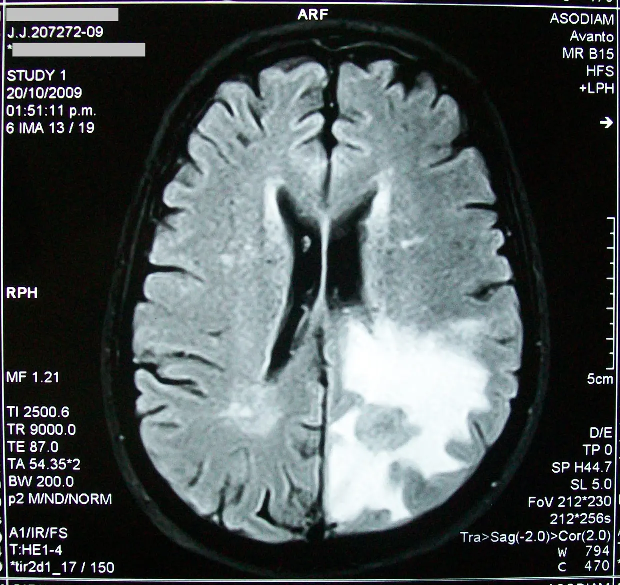
Researchers have reported a manganese-doped near-infrared quantum dot that combines fluorescence imaging with magnetic resonance imaging (MRI) in traumatic brain injury (TBI), according to a study in Science Advances. science.org
Science Advancesscience.orgOpen source ↗ The probe targets the limits of conventional imaging in showing the pathological events that determine disease progression and outcome.
The authors combined whole-brain clearing and cellular-resolution three-dimensional imaging with in vitro studies on different cell types involved in TBI. After systemic administration, the nanoprobes left the vessels through the compromised blood-brain barrier. They were then selectively phagocytosed by activated microglia.
The microglia subsequently migrated into regions rich in dead neurons and neuronal debris. According to the authors, this microglia-mediated tropism explains the origin of the MRI signal enhanced by the nanometer-sized contrast agent, turning MRI from an anatomical tool into a pathological reporter.
The team also compared the magnetic quantum dot with a biocompatible Fe3O4 nanoparticle of similar size, to assess the translational merits of nanoparticles for MRI diagnosis of TBI. science.org
Science Advancesscience.orgOpen source ↗ The work is at the laboratory stage; the material reports no clinical testing.
出典 · 1
誤りを見つけましたか?あわせて読みたい