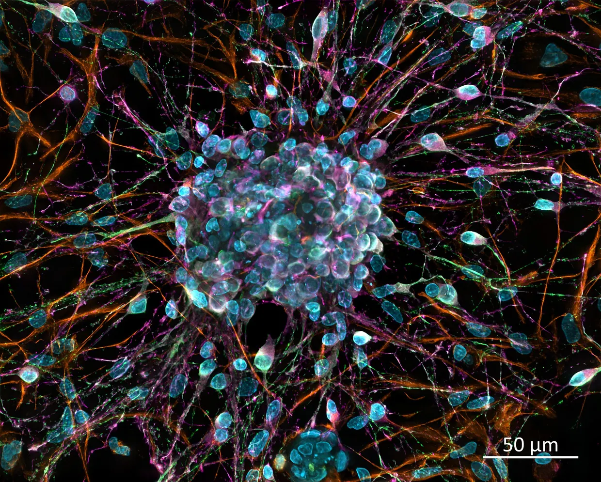
Chrm2 interneurons in the mouse hippocampus gate ripples and memory consolidation during sleep
Suppressing a sparse group of hippocampal interneurons during non-REM sleep impaired memory in mice, while ordinary sleep measures changed little.

A study in Neuron reports that a sparse population of GABAergic interneurons in the mouse hippocampus gates ripple generation and memory consolidation during sleep sciencedirect.com
Neuronsciencedirect.comOpen source ↗. The cells are identified by expression of the M2 acetylcholine receptor (Chrm2). The work was done in mice.
Cells active in non-REM sleep
According to the authors, all known genetically labeled hippocampal interneurons were highly active during wakefulness and REM sleep. Targeting Chrm2-expressing neurons isolated a distinct population, preferentially active during non-REM (NREM) sleep and quiet wakefulness. Acetylcholine inhibits these cells. The transmitter is more abundant during active wakefulness and REM sleep and falls during NREM sleep, which reduces its braking effect on them. The cells inhibited the entire hippocampus in an infra-slow rhythm of about 0.015 Hz.
Ripples are brief bursts of electrical activity that the hippocampus produces at rest. They are thought to help stabilize new memories. The authors report that the activity of Chrm2+ neurons modulated ripple occurrence in both directions through widespread inhibition. Stimulating the cells during NREM sleep made ripples more frequent, and quieting them made ripples less frequent.
Memory tests in mice
In an object-location test, mice explored two identical objects and then returned to their cages for five hours, during which they mostly slept. When they came back, one object had been moved. Control mice spent more time investigating the moved object, which suggests they remembered the earlier arrangement. Mice whose targeted cells were suppressed after learning showed little preference.
The researchers then used light to reduce the activity of these cells at different times. Suppression during NREM sleep made later recall worse. Suppression during wakefulness or REM sleep had no clear effect in this experiment. Ordinary measures of sleep showed little change. Suppressing the cells in dorsal CA3 impaired spatial memory, and suppressing them in the ventral hippocampus impaired social memory sciencedirect.com
Neuronsciencedirect.comOpen source ↗. A separate test suggested that related cells also helped mice recognize a mouse they had met before.
Existing human brain data show cells with a similar molecular marker. This gives researchers a place to look, but it does not establish that these cells have the same role in people. The study does not show how to diagnose memory problems from a sleep recording. It also leaves open how the cells work with their neighbors to produce ripples. The authors note that the cells offer genetically targeted access to sleep-active memory circuits implicated in cognitive disorders.