Two amyloid-β intermediates with distinct toxicity found in mice and human Alzheimer's brain tissue
Authors combined super-resolution microscopy with single-molecule imaging to classify amyloid-β intermediates by membrane damage; only one type converts into plaques.
A research team reported in Nature Communications two functionally distinct amyloid-β (Aβ) intermediates. They were found in recombinant Aβ42, in an Alzheimer's knock-in mouse model and in postmortem human Alzheimer's brain tissue. The authors combined super-resolution microscopy and single-molecule functional imaging with kinetic modelling. They classified the intermediates by their membrane-disrupting activity and linked this activity to the aggregation pathways that produce them nature.com
Nature Communicationsnature.comOpen source ↗.
Single-aggregate membrane assay
Alzheimer's disease affects over fifty million people worldwide. The authors note that plaque burden correlates poorly with cognitive decline and that plaque-clearing immunotherapies have shown only modest clinical benefit. Aβ aggregation cannot be monitored directly in the living human brain, and postmortem histology gives only static snapshots.
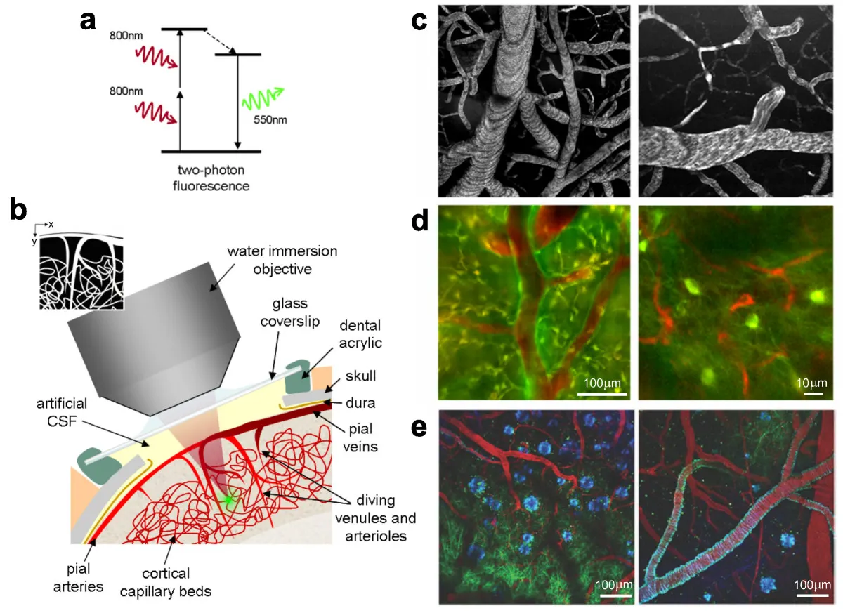
To address this, the team built a digital assay that detects individual Aβ species by their damaging potential. Nanoscale vesicles made of neuronal membrane-mimicking lipids were loaded with the dextran-conjugated Ca2+-sensitive dye Cal-520. The vesicles were immobilised on a coverslip in a Ca2+-containing buffer. If an aggregate permeabilises a vesicle, external Ca2+ enters and the dye fluoresces.
The signal was recorded from hundreds of vesicles at once by total internal reflection fluorescence (TIRF) microscopy. Each vesicle has an attoliter-scale volume (10−18 L), and concentrations were chosen so that at most one aggregate met a vesicle at a time. According to the authors, the assay registers events even when triggered by the passage of a single Ca2+ ion nature.com
Nature Communicationsnature.comOpen source ↗.
Two intermediate populations
Two intermediate populations were present in all three systems. They form through separate aggregation routes and differ in neurotoxicity, abundance and susceptibility to inhibition. Both damage neurons and glia, but only one converts into plaques.
In mice, pharmacological suppression of these intermediates reduced plaque burden, attenuated astrogliosis and improved cognition. The human data come from postmortem tissue. The authors conclude that the pathways of intermediate formation can serve as therapeutic targets in protein-misfolding disorders.
Fuentes · 1
¿Has encontrado un error?Lee también