Tübingen University Hospital performs first STAR procedure for ventricular tachycardia
Physicians treated a patient with life-threatening cardiac arrhythmia using non-invasive stereotactic arrhythmia radioablation.
In early October, specialists at the Tübingen University Hospital successfully treated a patient suffering from life-threatening ventricular tachycardia using targeted cardiac radiation, lomazoma.com reported lomazoma.com
lomazoma.comlomazoma.comOpen source ↗. The procedure, known as stereotactic arrhythmia radioablation (STAR), was implemented for the first time at the medical center by a multidisciplinary team combining radiation oncology and interventional cardiology. Following the non-invasive intervention, the patient's condition improved rapidly, and no further arrhythmias were recorded.
Ventricular tachycardia causes rapid ventricular contractions that can induce unconsciousness or sudden cardiac death, especially in individuals with preexisting myocardial damage. Standard interventions typically involve antiarrhythmic medication and invasive catheter ablation to eliminate defective cardiac tissue. However, a subset of patients does not respond adequately to these standard approaches, leaving recurrent episodes unmanaged.
Targeted radiotherapy protocol
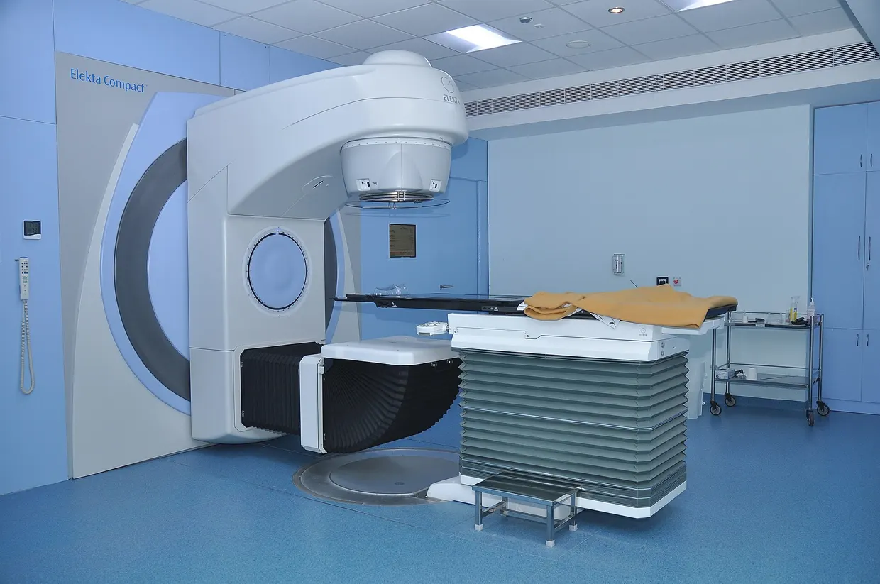
The STAR technique serves as a treatment option for selected patients when catheter-based and pharmacological interventions fail. The actual irradiation takes only a few minutes and does not require general anesthesia. During the procedure, linear accelerators deliver a single high-precision radiation dose externally to the exact myocardial zone generating the abnormal electrical signals.
Multimodal preparation and planning
The preparation for STAR requires extensive diagnostic mapping across multiple departments lomazoma.com
lomazoma.comlomazoma.comOpen source ↗. Electrophysiologists first isolate the arrhythmogenic substrates using cardiac catheter examinations. These electrophysiological datasets are subsequently merged with radiological imaging.
Using these combined models, medical physicists calculate an individualized radiation plan that covers the target volume while sparing surrounding healthy structures such as the lungs, esophagus, and stomach. The planning followed guidelines defined by the European research network STOPSTORM.