Shanghai Jiao Tong team maps finger flexor compartments in 3D with ultrasound and MRI in 10 people
Dynamic ultrasound plus MRI segmented compartments of the flexor digitorum superficialis in vivo and gave 3D fiber trajectories; the study involved ten healthy people.
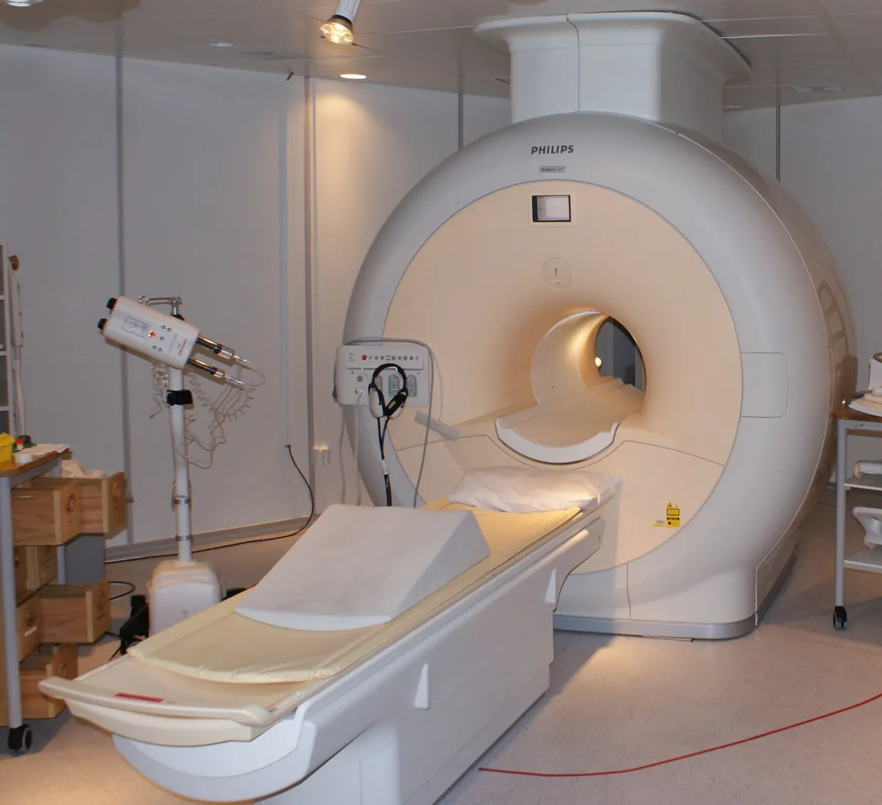
Researchers from Shanghai Jiao Tong University, Imperial College London and Siemens Healthineers in Shanghai have described a method for mapping neuromuscular compartments of the flexor digitorum superficialis in living humans. The work, published in Nature Communications on 3 November 2026, combines ultrasound and magnetic resonance imaging (MRI) and was tested in ten healthy individuals. nature.com
Nature Communicationsnature.comOpen source ↗
Neuromuscular compartments are the functional substrate of finger-specific motor control. Their three-dimensional (3D) architecture in vivo had remained unresolved, because imaging offers no clear fascial landmarks for them.
Ultrasound–MRI segmentation
The method uses two measurements of the same person. During isolated finger flexion, the authors recorded dynamic ultrasound and estimated the directions of tissue movement. According to the paper, these directions delineate the compartment contours.
The contours are then mapped into a 3D MRI reference, which produces volumetric compartment masks. The masks serve as constraints for tractography. It reconstructs 3D fiber trajectories and yields architectural measurements specific to each compartment. nature.com
Nature Communicationsnature.comOpen source ↗
Comparison with anatomy and electromyography
In the ten participants, compartment topology and fiber orientations were consistent with classic cadaveric anatomy, the authors report. The estimated compartments also accurately predicted the distributions of surface electromyography amplitude.
The reported data come from healthy volunteers and cover a single muscle, the flexor digitorum superficialis.
According to the authors, the framework provides 3D fiber trajectories and quantitative parameters for finger-specific computational models. They describe it as a foundation for subject-specific neuromuscular digital twins and next-generation neuromuscular interfaces. nature.com
Nature Communicationsnature.comOpen source ↗