BNITM and UKE: blocking RIPK1 in mice prolongs survival in cerebral malaria
Hamburg researchers found that the protein RIPK1 contributes to damage of brain vascular cells in malaria-infected mice. Human tissue and genetic data point to it as well.
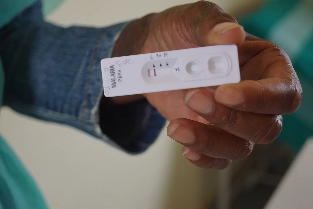
On 9 November 2026, researchers from the Bernhard Nocht Institute for Tropical Medicine (BNITM) and the University Medical Center Hamburg-Eppendorf (UKE), together with international partners, reported in Science Advances that the protein RIPK1 contributes to damage of vascular cells in the brains of malaria-infected mice, according to lomazoma.com lomazoma.com
lomazoma.comlomazoma.comOpen source ↗. RIPK1 regulates inflammation and cell death. Human brain tissue and genetic data from malaria patients also pointed to this protein.
WHO estimates for 2024 give over 280 million malaria cases and 610,000 deaths, 95 percent of them in sub-Saharan Africa, with children under five most affected. Cerebral malaria is marked by deep coma. About 25 to 30 percent of children who survive it remain impaired in motor skills, learning or memory.
How the team traced RIPK1
The researchers infected mice with Plasmodium berghei and analyzed messenger RNAs bound to the protein-making machinery of endothelial cells. Gene programs linked to cell death became increasingly active. CD8-positive T cells contribute to vascular damage in this model, and removing them weakened the cell-death programs in endothelial cells. Brain sections of infected mice showed raised markers of several cell-death processes.
In tissue from eight children in Malawi who died of cerebral malaria, endothelial cells showed stronger signals of activated RIPK1 than in eight children who died of other severe brain diseases without malaria lomazoma.com
lomazoma.comlomazoma.comOpen source ↗. This does not show whether CD8-positive T cells are causally linked to RIPK1 activation. Existing genetic data, covering several forms of severe malaria and not only the cerebral form, revealed associations between variants of regulated cell-death genes, including the RIPK1 signaling network, and severe malaria. The authors note that this does not prove the disease mechanism runs through RIPK1.
Longer survival in the mouse model
In genetically modified mice, the enzymatic activity of RIPK1 was switched off specifically in brain endothelial cells. After infection these mice lived longer than controls, but the change did not prevent death. In a separate group, the team inhibited RIPK1 with the experimental compound GNE684, which can also act outside brain vessels. Treatment began on the first day after infection, before neurological disease appeared, and these mice also survived longer. Parasite load in the blood stayed comparable to controls in both groups.
The mouse model reflects important aspects of vascular damage but differs from human malaria in key disease mechanisms. Whether RIPK1 inhibition improves survival or prevents later neurological damage in patients remains open and requires further study. The next step is to test whether later administration alongside standard malaria treatment is effective and tolerable.
Sources · 1
Vous avez trouvé une erreur ?À lire aussi