In SCA17 models, calpain cleaves TBP after T106 and alters neuronal AMOT–YAP1 signalling
Authors mapped a main calpain cleavage site in TBP, the protein mutated in spinocerebellar ataxia type 17, and linked its fragments to AMOT; tested in cells, rat and zebrafish.
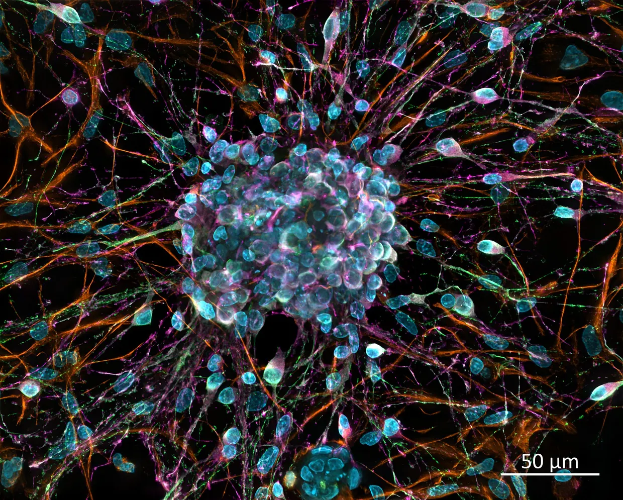
A research team reported in Nature Communications that calpain, a calcium-activated protease, cleaves the TATA-box-binding protein (TBP) mainly after residue T106, just C-terminal to its polyglutamine (polyQ) tract nature.com
Nature Communicationsnature.comOpen source ↗. Polyglutamine expansion in TBP causes spinocerebellar ataxia type 17 (SCA17). The authors worked with SCA17 cell lines, patient-derived fibroblasts and induced neurons, and with rat and zebrafish models.
TBP, SCA17 and the cleavage site
TBP is an essential subunit of the TFIID complex. Its conserved C-terminal core domain recognises TATA-box motifs and helps recruit RNA polymerase II. Normal TBP alleles contain 25–40 repeats, pathogenic alleles more than 41, with full penetrance at 50. SCA17 is an autosomal dominant disorder with ataxia, dementia, involuntary movements and psychiatric symptoms, and the mechanisms of neuronal loss remain unclear.
In vitro, myc-tagged full-length TBP with 38Q or 106Q was overexpressed in HEK 293T cells, immunopurified and cleaved with calpain-1. Western blotting showed three C-terminal fragments (c1–c3) and three polyQ-containing N-terminal fragments (n1–n3). The antibody D5G7Y, which binds around residue A110, detected only fragment c1, so that cleavage lies N-terminal to the epitope. In cells co-expressing TBP and calpain-1, TBP was mainly nuclear, while calpain-1 was found only in the cytoplasm.
Fragments, calpain and AMOT
According to the authors, the polyQ-containing N-terminal fragments mislocalise to the cytoplasm and aggregate more. Both the N-terminal and the nuclear C-terminal fragments contribute to calpain overactivation nature.com
Nature Communicationsnature.comOpen source ↗.
Multi-omics analyses in an SCA17 cell line showed that the TBP fragments alter pathways linked to neuronal function and neurodegeneration, with AMOT identified as a key player. Further experiments in cell lines, patient-derived fibroblasts and induced neurons, as well as in rat and zebrafish models, confirmed a role of AMOT in neurons. The authors report that AMOT is regulated by TBP and acts via YAP1-dependent signalling.
The authors state that the findings add to the understanding of TBP's neuronal function and point to wider relevance of AMOT in neurodegenerative and neurodevelopmental disorders. The data come from cellular and animal models and from patient-derived cells.
Quellen · 1
Einen Fehler gefunden?Lesen Sie auch